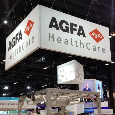
Agfa HealthCare plans to showcase products in its artificial intelligence (AI) and digital radiography (DR) portfolio at France's national radiology congress (JFR), which begins on 11 October in Paris.
Company highlights include the Musica Nerve Center acquisition workstation, which is powered by AI and deep learning for enhanced operational efficiency and features a single, intuitive interface for DR imaging.
Agfa's DR systems range from entry-level mobile units to advanced, multipurpose, automated x-ray rooms, designed for enhanced image quality and low radiation dose exposure to patients and staff. The mobile DR 100e can be moved down narrow corridors for both bedside and intensive care unit (ICU) imaging. It also features Musica image processing software for improved productivity.
Also at JFR 2019 will be SE Suite, a diagnostic multimodality workstation for the manipulation, archiving, and distribution of medical imaging data. It offers an intuitive screen layout for navigation between images and studies, and the workstation can be tailored for single- or multiuser environments, including those that require a specific workflow, such as mammography screening and orthopedics. according to the vendor.




![A normal mammogram confirmed by three-year radiologic follow-up illustrates reader-marked regions of interest (ROIs) during (A) unaided (round 1) and (B) artificial intelligence (AI)–assisted (round 2) reading. Each colored dot represents an ROI for recall by a human reader. Readers could mark more than one ROI per case, represented by multiple dots of the same color. During AI-assisted reading, the AI system displayed three visible prompts: two with suspicion of malignancy scores of 35% (left mediolateral oblique [L MLO] and craniocaudal [L CC]) and one with a suspicion of malignancy score of 10% (right craniocaudal [R CC]), shown as polygonal overlays. Without AI, six of 10 readers (60%) marked a false-positive ROI. With AI assistance, this fell to two of 10 (20%). R MLO = right mediolateral oblique.](https://img.auntminnieeurope.com/mindful/smg/workspaces/default/uploads/2026/07/2026-07-14-radiology-mammogram-ai-auto-bias.H0bYO8QlWs.jpg?auto=format%2Ccompress&fit=crop&h=100&q=70&w=100)


![A normal mammogram confirmed by three-year radiologic follow-up illustrates reader-marked regions of interest (ROIs) during (A) unaided (round 1) and (B) artificial intelligence (AI)–assisted (round 2) reading. Each colored dot represents an ROI for recall by a human reader. Readers could mark more than one ROI per case, represented by multiple dots of the same color. During AI-assisted reading, the AI system displayed three visible prompts: two with suspicion of malignancy scores of 35% (left mediolateral oblique [L MLO] and craniocaudal [L CC]) and one with a suspicion of malignancy score of 10% (right craniocaudal [R CC]), shown as polygonal overlays. Without AI, six of 10 readers (60%) marked a false-positive ROI. With AI assistance, this fell to two of 10 (20%). R MLO = right mediolateral oblique.](https://img.auntminnieeurope.com/mindful/smg/workspaces/default/uploads/2026/07/2026-07-14-radiology-mammogram-ai-auto-bias.H0bYO8QlWs.jpg?auto=format%2Ccompress&fit=crop&h=112&q=70&w=112)