The French national radiology congress (JFR) kicked off officially on Friday with a strengthened focus on artificial intelligence (AI) and machine learning. The discussion included an update on an ambitious project to create a national ecosystem in France for AI algorithm development.
Speaking at the AI teaching session "AI: What dynamics in 2019?" Dr. Jean-Paul Beregi, head of imaging at Nȋmes university hospital, updated delegates on the current status of France's new DRIM France IA imaging initiative, a nonprofit, independent AI ecosystem.
The project has advanced significantly since it was announced last October, according to Beregi. The ecosystem now has a scientific committee and a charter, and is looking at how to move forward with institutional partners.
"We are now at the stage of being able to assess the roles and contributions of other actors," Beregi noted.
It is hoped that DRIM France will be sufficiently established to serve as a neutral testing platform for private startups or public hospitals wanting to validate their algorithms on a concrete database as early as 2020, according to Beregi.
In addition, DRIM France is likely to play a more major role in next year's Big Data Challenge at JFR 2020, with the aim of expanding the competition to include AI's impact on questions relating not just to pathology but also to dose, pertinence, and prognosis. Teams will have access to other nonimage information on clinical indication, biology, dose, and patient history taken from RIS and PACS.
The current JFR data challenge event starts today with 18 teams. They have until Sunday evening to submit their findings using their own algorithms. This year the database contains 4,500 CT and MR images across the three topics of lung nodules, multiple sclerosis, and sarcopenia.




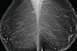
![A normal mammogram confirmed by three-year radiologic follow-up illustrates reader-marked regions of interest (ROIs) during (A) unaided (round 1) and (B) artificial intelligence (AI)–assisted (round 2) reading. Each colored dot represents an ROI for recall by a human reader. Readers could mark more than one ROI per case, represented by multiple dots of the same color. During AI-assisted reading, the AI system displayed three visible prompts: two with suspicion of malignancy scores of 35% (left mediolateral oblique [L MLO] and craniocaudal [L CC]) and one with a suspicion of malignancy score of 10% (right craniocaudal [R CC]), shown as polygonal overlays. Without AI, six of 10 readers (60%) marked a false-positive ROI. With AI assistance, this fell to two of 10 (20%). R MLO = right mediolateral oblique.](https://img.auntminnieeurope.com/mindful/smg/workspaces/default/uploads/2026/07/2026-07-14-radiology-mammogram-ai-auto-bias.H0bYO8QlWs.jpg?auto=format%2Ccompress&fit=crop&h=100&q=70&w=100)


![A normal mammogram confirmed by three-year radiologic follow-up illustrates reader-marked regions of interest (ROIs) during (A) unaided (round 1) and (B) artificial intelligence (AI)–assisted (round 2) reading. Each colored dot represents an ROI for recall by a human reader. Readers could mark more than one ROI per case, represented by multiple dots of the same color. During AI-assisted reading, the AI system displayed three visible prompts: two with suspicion of malignancy scores of 35% (left mediolateral oblique [L MLO] and craniocaudal [L CC]) and one with a suspicion of malignancy score of 10% (right craniocaudal [R CC]), shown as polygonal overlays. Without AI, six of 10 readers (60%) marked a false-positive ROI. With AI assistance, this fell to two of 10 (20%). R MLO = right mediolateral oblique.](https://img.auntminnieeurope.com/mindful/smg/workspaces/default/uploads/2026/07/2026-07-14-radiology-mammogram-ai-auto-bias.H0bYO8QlWs.jpg?auto=format%2Ccompress&fit=crop&h=112&q=70&w=112)