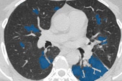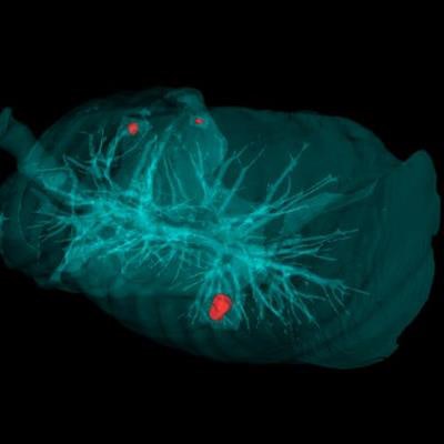
Researchers from Russia have developed an artificial intelligence (AI) program called Doctor AI-zimov that is capable of identifying and marking lung nodules -- some as small as 2 mm -- on CT scans in under 20 seconds.
The AI software spots lung nodules on segmented CT scans and uses a recently patented "chord" method to connect a series of points randomly drawn onto each nodule. Next, the software situates the nodule in a virtual cube and composes perpendicular lines from the edges of the cube to the surface of the nodule. This process results in the generation of simple histograms that reflect the shape and structure of the nodules.
Finally, an algorithm analyzes these histograms to determine the size and potential malignancy of each nodule and then sends the results to clinicians for review.
 The AI software can flag lung nodules on CT scans within 20 seconds. Image courtesy of Peter the Great St. Petersburg Polytechnic University.
In terms of computing file size, the histograms are considerably smaller than the average CT scan, which allows the algorithm to complete tasks within seconds, lead researcher Lev Utkin, PhD, from Peter the Great St. Petersburg Polytechnic University said in a statement. What's more, the AI software is capable of detecting lung nodules as small as 2 mm.
"Initially, we set up an algorithm to search for nodules starting from 6 mm, because radiologists themselves start the treatment of tumors of this size," Utkin said. "But the system is so smart that it was able to find nodules of even smaller size."
Utkin and colleagues trained the software on 1,000 CT scans from the Lung Image Database Consortium (LIDC). They also tested the software on the CT scans of 60 patients at their institution's oncology center and have obtained favorable results.
The researchers plan to continue open testing of the software at the St. Petersburg Clinical Research Center. One of their long-term goals is to adapt the software to be able to analyze x-rays and ultrasound scans of various organs.