Royal Philips, the parent company of Philips Healthcare, is highlighting results from a global study indicating that automated 3D analysis of echocardiography images of the left heart chambers is an accurate alternative to conventional manual analysis.
Current guidelines recommend 3D echo chamber quantification for patients undergoing an echocardiography exam, but adoption in clinical practice has lagged due to time-consuming analysis.
This study compared Philips' HeartModelA.I. technique with a conventional manual method to determine the accuracy and reproducibility of three different heart measurements in a multicenter setting: left-atrial (LA) volume, left-ventricular (LV) volume, and ejection fraction. The research team found the Philips technique to be just as accurate as the conventional method (EHJ - Cardiovascular Imaging, 4 February 2017).
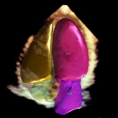
Philips HeartModelA.I. provides automated 3D analysis of echocardiography images. Image courtesy of Philips.Lead author Dr. Diego Medvedofsky and colleagues imaged 180 patients across six sites. Images were analyzed using the automated HeartModelA.I. software with endocardial border correction when necessary and by manual tracing. Measurements were performed by each site and by the core laboratory as the reference.
Comparisons without corrections showed perfect agreement for all parameters. With corrections, correlations were better, demonstrating the automated 3D echo technique to be just as accurate.




![A normal mammogram confirmed by three-year radiologic follow-up illustrates reader-marked regions of interest (ROIs) during (A) unaided (round 1) and (B) artificial intelligence (AI)–assisted (round 2) reading. Each colored dot represents an ROI for recall by a human reader. Readers could mark more than one ROI per case, represented by multiple dots of the same color. During AI-assisted reading, the AI system displayed three visible prompts: two with suspicion of malignancy scores of 35% (left mediolateral oblique [L MLO] and craniocaudal [L CC]) and one with a suspicion of malignancy score of 10% (right craniocaudal [R CC]), shown as polygonal overlays. Without AI, six of 10 readers (60%) marked a false-positive ROI. With AI assistance, this fell to two of 10 (20%). R MLO = right mediolateral oblique.](https://img.auntminnieeurope.com/mindful/smg/workspaces/default/uploads/2026/07/2026-07-14-radiology-mammogram-ai-auto-bias.H0bYO8QlWs.jpg?auto=format%2Ccompress&fit=crop&h=100&q=70&w=100)


![A normal mammogram confirmed by three-year radiologic follow-up illustrates reader-marked regions of interest (ROIs) during (A) unaided (round 1) and (B) artificial intelligence (AI)–assisted (round 2) reading. Each colored dot represents an ROI for recall by a human reader. Readers could mark more than one ROI per case, represented by multiple dots of the same color. During AI-assisted reading, the AI system displayed three visible prompts: two with suspicion of malignancy scores of 35% (left mediolateral oblique [L MLO] and craniocaudal [L CC]) and one with a suspicion of malignancy score of 10% (right craniocaudal [R CC]), shown as polygonal overlays. Without AI, six of 10 readers (60%) marked a false-positive ROI. With AI assistance, this fell to two of 10 (20%). R MLO = right mediolateral oblique.](https://img.auntminnieeurope.com/mindful/smg/workspaces/default/uploads/2026/07/2026-07-14-radiology-mammogram-ai-auto-bias.H0bYO8QlWs.jpg?auto=format%2Ccompress&fit=crop&h=112&q=70&w=112)