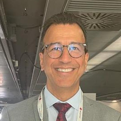
Comment les secteurs public et privé travaillent-ils ensemble dans la radiologie française? Le professeur Afshin Gangi, de Strasbourg, nous éclaire sur le système unique de la France. Dans une interview vidéo réalisée par notre correspondante Mélisande Rouger à l'occasion des Journées nationales de radiologie (JFR 2022), il évoque également comment son équipe évite le burnout.
"La France est très différente des autres pays. Les hôpitaux publics sont contrôlés à 100 % par le gouvernement français," explique M. Gangi, qui est chef du service de radiologie de l'hôpital universitaire de Strasbourg et professeur au King's College de Londres. Les radiologues peuvent toutefois travailler à la fois dans le secteur public et dans le secteur privé s'ils consacrent au moins 20 % de leur temps à l'autre secteur, a-t-il ajouté.
Bien qu'il ne ressente aucune tension entre les radiologues publics et privés, Gangi reconnaît qu'un certain "parisianisme" -- une tendance à séparer Paris du reste de la France - persiste en radiologie et dans les soins de santé en général. "Le parisianisme est inévitable en France," a-t-il déclaré. "C'est un pays assez centralisé. Mais même dans notre système universitaire national, il y a beaucoup de gens qui viennent de l'extérieur de la capitale."
Il n'y a pas assez de radiologues interventionnels en France, selon Gangi, qui est le président sortant de la Société européenne de radiologie cardiovasculaire et interventionnelle (CIRSE). "Dans mon équipe, chacun d'entre nous travaille un jour par semaine dans deux autres hôpitaux à Colmar et à Mulhouse," a-t-il déclaré. "Ce n'est pas suffisant pour les 2 millions d'habitants que nous devons couvrir. Il nous manque plus de 100 à 200 radiologues interventionnels en France."
La radiologie interventionnelle reste une sous-spécialité relativement dominée par les hommes et des mesures doivent être prises pour améliorer la situation, a-t-il concédé. "65 % des étudiants en médecine sont des femmes," a noté Gangi. "Pour votre avenir, vous devez faire quelque chose pour survivre."
Le burnout est un problème en radiologie en général, mais heureusement pas dans son groupe, estime-t-il. "Mon équipe est composée de pilotes fous, cela signifie que nous sommes toujours en train de faire la course! Nous avons de la chance, nous avons beaucoup de nouveautés," a-t-il déclaré. "Personne n'est touché par le burnout, mais je reconnais le problème à l'hôpital. Le burnout tue la motivation de certaines personnes. Le COVID a rendu ce phénomène encore plus évident."
L'enthousiasme est son remède préventif. "Quand les patients vous voient après le traitement et qu'ils sont heureux, cela recharge votre batterie," a-t-il conclu.
Le professeur Afshin Gangi a évoqué la structure la radiologie en France -- ainsi que le phénomène du "parisianisme" -- lors du congrès national français de radiologie, les JFR 2022.
Note de rédacteur: Ceci est une traduction éditée d'un article publié en anglais en ligne par AuntMinnieEurope. Traduction par Frances Rylands-Monk.


![Examples of ultrasound findings and techniques. (A) Images in a 39-year-old male patient with a mass in the left thigh. The mass is heterogeneous on the B-mode US image (compared with the patient in D) and showed increased microvascularity (superb microvascular imaging [SMI]) and shear-wave elastography (SWE) values. Undifferentiated pleomorphic sarcoma was diagnosed at biopsy (with pleomorphic rhabdomyosarcoma in surgical specimen). (B) Images in an 18-year-old male patient with a mass in the left leg. The mass is hypoechoic on the B-mode image, with no other findings suggestive of malignancy. The lesion is in contact with the cortex of the tibia, which is slightly irregular. CT revealed a doubtful anteromedial tibial erosion. The microvascular study demonstrated high vascularization, suggestive of malignancy. Periosteal Ewing sarcoma was diagnosed with both histologic and immunohistochemical confirmation. (C) Images in a 69-year-old female patient with a lump growing on the outside of the left leg. Multiple SWE examinations were performed (please note the high values obtained in the measurements, whereas the color map highlights the stiffness relative to adjacent tissues). SMI showed areas of increased vascularization to target for sampling. Undifferentiated spindle cell sarcoma was diagnosed at biopsy, with residual leiomyosarcoma in the surgical specimen after neoadjuvant therapy. (D) Images in a 56-year-old female patient with a mass in the right thigh. The mass is heterogeneous at both B-mode ultrasound (similar to patient A) and MRI (coronal T2-weighted spectral attenuated inversion recovery [SPAIR]; T1-weighted pre-contrast and postcontrast imaging), which even shows uptake after the administration of paramagnetic contrast material, which is traditionally suggestive of malignancy. Low values at SMI and elastography are suggestive of benignity. Spindle cell lipoma was diagnosed at biopsy, with atypical spindle cell lipomatous tumor in the surgical specimen.](https://img.auntminnieeurope.com/mindful/smg/workspaces/default/uploads/2026/08/images-radiol250278fig2.APCFLSvX6p.jpg?auto=format%2Ccompress&fit=crop&h=100&q=70&w=100)


![Examples of ultrasound findings and techniques. (A) Images in a 39-year-old male patient with a mass in the left thigh. The mass is heterogeneous on the B-mode US image (compared with the patient in D) and showed increased microvascularity (superb microvascular imaging [SMI]) and shear-wave elastography (SWE) values. Undifferentiated pleomorphic sarcoma was diagnosed at biopsy (with pleomorphic rhabdomyosarcoma in surgical specimen). (B) Images in an 18-year-old male patient with a mass in the left leg. The mass is hypoechoic on the B-mode image, with no other findings suggestive of malignancy. The lesion is in contact with the cortex of the tibia, which is slightly irregular. CT revealed a doubtful anteromedial tibial erosion. The microvascular study demonstrated high vascularization, suggestive of malignancy. Periosteal Ewing sarcoma was diagnosed with both histologic and immunohistochemical confirmation. (C) Images in a 69-year-old female patient with a lump growing on the outside of the left leg. Multiple SWE examinations were performed (please note the high values obtained in the measurements, whereas the color map highlights the stiffness relative to adjacent tissues). SMI showed areas of increased vascularization to target for sampling. Undifferentiated spindle cell sarcoma was diagnosed at biopsy, with residual leiomyosarcoma in the surgical specimen after neoadjuvant therapy. (D) Images in a 56-year-old female patient with a mass in the right thigh. The mass is heterogeneous at both B-mode ultrasound (similar to patient A) and MRI (coronal T2-weighted spectral attenuated inversion recovery [SPAIR]; T1-weighted pre-contrast and postcontrast imaging), which even shows uptake after the administration of paramagnetic contrast material, which is traditionally suggestive of malignancy. Low values at SMI and elastography are suggestive of benignity. Spindle cell lipoma was diagnosed at biopsy, with atypical spindle cell lipomatous tumor in the surgical specimen.](https://img.auntminnieeurope.com/mindful/smg/workspaces/default/uploads/2026/08/images-radiol250278fig2.APCFLSvX6p.jpg?auto=format%2Ccompress&fit=crop&h=112&q=70&w=112)



