CHICAGO - If and when it is ready for routine clinical use, 7-tesla time-of-flight (TOF) MR angiography (MRA) will provide "superior assessment" of aneurysms and related vessel features, according to a study presented on Tuesday at RSNA 2013.
The findings from German researchers at Essen University Hospital validate the use of 7-tesla TOF MRA to overcome limitations of 1.5-tesla imaging for assessing intracranial aneurysms, due to its high-quality vessel-tissue contrast and improved spatial resolution.
Ruptured intracranial aneurysms are considered the main cause of subarachnoid hemorrhage, so detection and assessment of aneurysm localization and related features are critical for appropriate treatment planning, the researchers noted.
Ultrahigh-field MRA "may be particularly important for patients with small aneurysms," said lead author Dr. Lale Umutlu, from the university's Institute of Diagnostic and Interventional Radiology and Neuroradiology. However, while 7-tesla MRI in general will remain "an important focus of scientific research, the step to the clinical routine will probably be rather restricted," she added.
Limited detection
The problem with 1.5-tesla MRI is that it's limited in the detection of small aneurysms. Ultrahigh-field MRA can provide more detailed images and a better assessment of a patient's intracranial vasculature based on higher spatial resolution from the increased signal-to-noise ratio.
"The increase of the magnetic field strength to 7 tesla is associated with a significant increase of the signal-to-noise ratio that can be transitioned into imaging at higher spatial or temporal resolution," Umutlu told AuntMinnie.com.
For the current study, 17 subjects were examined using 1.5-tesla TOF MRA (Magnetom Aera, Siemens Healthcare), followed by the 7-tesla TOF MRA scan (Magnetom 7T, Siemens).
Two radiologists assessed the two sets of MR angiograms for the aneurysm dome, neck, parent vessel, vessel tissue contrast, and image impairment due to any artifacts. They rated the images on a five-point scale, with 5 as "excellent delineation" and 1 as "nondiagnostic." Contrast ratios of all aneurysms and adjacent parenchyma also were calculated.
The researchers found that 7-tesla TOF MRA provided significantly superior delineation of the dome and neck, compared with 1.5-tesla MRA.
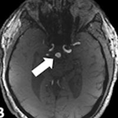
Image A shows 1.5-tesla TOF MRA of a 55-year-old patient with an unruptured basilar aneurysm (arrow). Image B is a corresponding 7-tesla TOF MRA and image C is a maximum intensity projection image. 7-tesla MRA produced superior delineation of the aneurysm with enhanced image sharpness and clarity. Images courtesy of Dr. Lale Umutlu."In our study, we were able to improve the spatial resolution to a minimum voxel size of 0.2 x 0.2 x 0.4 mm3 at 7 tesla, instead of 0.35 x 0.35 x 0.7 mm3 at 1.5 tesla," Umutlu explained. "Based on this superior spatial resolution, as well the improved vessel-to-tissue contrast, 7-tesla TOF MRA allowed for a significantly improved delineation of the intracranial aneurysms."
"Current studies focus on the comparison of TOF MRA to [digital subtraction angiography (DSA)], revealing comparable diagnostic accuracy for 7-tesla TOF MRA and DSA," she said. "Thus, follow-up of patients with unruptured aneurysms may be performed via 7-tesla TOF MRA, sparing the application of ionizing radiation and contrast agent."
Umutlu and colleagues plan to further investigate their findings and focus on the comparison of 7-tesla TOF MRA with DSA in patients with unruptured aneurysms, as well as arteriovenous malformations.


![Examples of ultrasound findings and techniques. (A) Images in a 39-year-old male patient with a mass in the left thigh. The mass is heterogeneous on the B-mode US image (compared with the patient in D) and showed increased microvascularity (superb microvascular imaging [SMI]) and shear-wave elastography (SWE) values. Undifferentiated pleomorphic sarcoma was diagnosed at biopsy (with pleomorphic rhabdomyosarcoma in surgical specimen). (B) Images in an 18-year-old male patient with a mass in the left leg. The mass is hypoechoic on the B-mode image, with no other findings suggestive of malignancy. The lesion is in contact with the cortex of the tibia, which is slightly irregular. CT revealed a doubtful anteromedial tibial erosion. The microvascular study demonstrated high vascularization, suggestive of malignancy. Periosteal Ewing sarcoma was diagnosed with both histologic and immunohistochemical confirmation. (C) Images in a 69-year-old female patient with a lump growing on the outside of the left leg. Multiple SWE examinations were performed (please note the high values obtained in the measurements, whereas the color map highlights the stiffness relative to adjacent tissues). SMI showed areas of increased vascularization to target for sampling. Undifferentiated spindle cell sarcoma was diagnosed at biopsy, with residual leiomyosarcoma in the surgical specimen after neoadjuvant therapy. (D) Images in a 56-year-old female patient with a mass in the right thigh. The mass is heterogeneous at both B-mode ultrasound (similar to patient A) and MRI (coronal T2-weighted spectral attenuated inversion recovery [SPAIR]; T1-weighted pre-contrast and postcontrast imaging), which even shows uptake after the administration of paramagnetic contrast material, which is traditionally suggestive of malignancy. Low values at SMI and elastography are suggestive of benignity. Spindle cell lipoma was diagnosed at biopsy, with atypical spindle cell lipomatous tumor in the surgical specimen.](https://img.auntminnieeurope.com/mindful/smg/workspaces/default/uploads/2026/08/images-radiol250278fig2.APCFLSvX6p.jpg?auto=format%2Ccompress&fit=crop&h=100&q=70&w=100)


![Examples of ultrasound findings and techniques. (A) Images in a 39-year-old male patient with a mass in the left thigh. The mass is heterogeneous on the B-mode US image (compared with the patient in D) and showed increased microvascularity (superb microvascular imaging [SMI]) and shear-wave elastography (SWE) values. Undifferentiated pleomorphic sarcoma was diagnosed at biopsy (with pleomorphic rhabdomyosarcoma in surgical specimen). (B) Images in an 18-year-old male patient with a mass in the left leg. The mass is hypoechoic on the B-mode image, with no other findings suggestive of malignancy. The lesion is in contact with the cortex of the tibia, which is slightly irregular. CT revealed a doubtful anteromedial tibial erosion. The microvascular study demonstrated high vascularization, suggestive of malignancy. Periosteal Ewing sarcoma was diagnosed with both histologic and immunohistochemical confirmation. (C) Images in a 69-year-old female patient with a lump growing on the outside of the left leg. Multiple SWE examinations were performed (please note the high values obtained in the measurements, whereas the color map highlights the stiffness relative to adjacent tissues). SMI showed areas of increased vascularization to target for sampling. Undifferentiated spindle cell sarcoma was diagnosed at biopsy, with residual leiomyosarcoma in the surgical specimen after neoadjuvant therapy. (D) Images in a 56-year-old female patient with a mass in the right thigh. The mass is heterogeneous at both B-mode ultrasound (similar to patient A) and MRI (coronal T2-weighted spectral attenuated inversion recovery [SPAIR]; T1-weighted pre-contrast and postcontrast imaging), which even shows uptake after the administration of paramagnetic contrast material, which is traditionally suggestive of malignancy. Low values at SMI and elastography are suggestive of benignity. Spindle cell lipoma was diagnosed at biopsy, with atypical spindle cell lipomatous tumor in the surgical specimen.](https://img.auntminnieeurope.com/mindful/smg/workspaces/default/uploads/2026/08/images-radiol250278fig2.APCFLSvX6p.jpg?auto=format%2Ccompress&fit=crop&h=112&q=70&w=112)



