Dear Women's Imaging Insider,
A team from Rome has pulled off a masterstroke: They've compiled an easy-to-follow and concise list of 10 reasons why you should care about breast AI.
Let's be honest, there's a lot of hype around right now about AI. There are many reasons to be optimistic, but serious implementation challenges remain. The strength of the approach taken by the Italian researchers is they're optimistic about the future -- yet also realistic about the difficult path to implementation. Get the full story in today's top article.
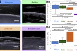
In another story posted this week, a French group has reported its latest findings on breast implant rupture. The team is convinced shear-wave elastography has a role to play in these cases.
Interestingly, Dutch investigators are convinced that 3D elastography can be applied to an automated breast volume scanner to analyze in vivo strain images.
Meanwhile, the authors of a paper published in the European Journal of Radiology have found that conebeam breast CT has superior diagnostic performance than that of mammography in small studies.
Looking ahead, we'll soon be bringing you all the important news from the European Congress of Radiology, which begins in Vienna on 28 February. Our buildup to the continent's premier medical imaging event will begin shortly. Watch this space!
Philip Ward
Editor in Chief
AuntMinnieEurope.com





![A normal mammogram confirmed by three-year radiologic follow-up illustrates reader-marked regions of interest (ROIs) during (A) unaided (round 1) and (B) artificial intelligence (AI)–assisted (round 2) reading. Each colored dot represents an ROI for recall by a human reader. Readers could mark more than one ROI per case, represented by multiple dots of the same color. During AI-assisted reading, the AI system displayed three visible prompts: two with suspicion of malignancy scores of 35% (left mediolateral oblique [L MLO] and craniocaudal [L CC]) and one with a suspicion of malignancy score of 10% (right craniocaudal [R CC]), shown as polygonal overlays. Without AI, six of 10 readers (60%) marked a false-positive ROI. With AI assistance, this fell to two of 10 (20%). R MLO = right mediolateral oblique.](https://img.auntminnieeurope.com/mindful/smg/workspaces/default/uploads/2026/07/2026-07-14-radiology-mammogram-ai-auto-bias.H0bYO8QlWs.jpg?auto=format%2Ccompress&fit=crop&h=100&q=70&w=100)


![A normal mammogram confirmed by three-year radiologic follow-up illustrates reader-marked regions of interest (ROIs) during (A) unaided (round 1) and (B) artificial intelligence (AI)–assisted (round 2) reading. Each colored dot represents an ROI for recall by a human reader. Readers could mark more than one ROI per case, represented by multiple dots of the same color. During AI-assisted reading, the AI system displayed three visible prompts: two with suspicion of malignancy scores of 35% (left mediolateral oblique [L MLO] and craniocaudal [L CC]) and one with a suspicion of malignancy score of 10% (right craniocaudal [R CC]), shown as polygonal overlays. Without AI, six of 10 readers (60%) marked a false-positive ROI. With AI assistance, this fell to two of 10 (20%). R MLO = right mediolateral oblique.](https://img.auntminnieeurope.com/mindful/smg/workspaces/default/uploads/2026/07/2026-07-14-radiology-mammogram-ai-auto-bias.H0bYO8QlWs.jpg?auto=format%2Ccompress&fit=crop&h=112&q=70&w=112)