Dear Women's Imaging Insider,
Current techniques for performing specimen radiography in patients undergoing breast-conserving surgery using full-field digital mammography have low sensitivities compared with final histopathological margin analysis. Could digital breast tomosynthesis be the answer breast imagers are looking for? Find out by clicking here.
In another article about breast-conserving surgery, researchers are reporting early success with the combination of optical and molecular imaging to view FDG-PET images that detect early-stage breast cancer. The technique, Cerenkov luminescence imaging, can assess tumor margins during breast tumor surgery by detecting light emitted from an FDG-PET radiopharmaceutical.
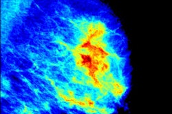
Also in your Women's Imaging Community, breast density software is getting reviewed. It's well established that spotting breast cancer in women with dense breasts is more challenging, so a study that targets breast density is particularly intriguing. A study from Spain found breast density software could improve reliability of density estimations within the radiology community, and it could open a window to start considering local breast distribution for risk assessment. Read more.
We also have two stories from the Middle East you won't want to miss. In the first, a new chart developed in Iran can help physicians identify women who are at high risk for breast cancer. Doing so could make a breast cancer screening program more efficient and productive. But are things as simple as that? Let us know in the Forums.
In the second Middle Eastern story, researchers from Saudi Arabia reported that MRI can confirm suspected placental anomalies or assist in cases of equivocal ultrasound findings. The study of pregnant women found that noncontrast MRI is a sensitive diagnostic test for detecting the increasingly common clinical problem of invasive placenta.
But wait! There's more. Be sure to head on over to the Women's Imaging Community to scope out the rest. And as always, I enjoy hearing from you, so contact me anytime.