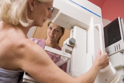
Musicians can suffer from a wide range of performance-related musculoskeletal and vocal disorders, including tendon and ligament injuries of the hand, wrist, and elbow; shoulder, neck, and spine disorders caused by prolonged posture and asymmetrical loading; nerve entrapments and overuse syndromes; vocal cord and laryngeal injuries in singers; foot and ankle problems in seated and standing performers; and impact and repetitive-strain injuries in percussionists.
We’ve published a new book (“Injuries in Musicians - Imaging and Management”) that examines these conditions through the lens of imaging, showing how injuries appear on MRI, ultrasound, and other modalities, and how imaging can guide both diagnosis and treatment.
 Sagittal T1-weighted (A) and STIR (B) MR images show small disc bulges and disc dehydration at multiple levels in keeping with degenerative change. All images courtesy of Prof. Rajesh Botchu.
This volume offers a dedicated, imaging-led, interdisciplinary reference guide for musician health. It speaks equally to radiologists, orthopedic surgeons, physiotherapists, and performing arts medicine specialists -- and it also gives musicians a clearer understanding of what is happening inside their bodies.
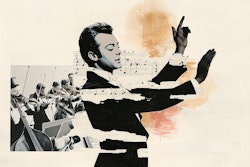
By combining cultural history, instrument science, radiology, orthopaedics, rehabilitation, and musical aesthetics, the book reflects the reality that musical performance is both an art and a physical discipline. That integrated vision is what motivated the editors -- and what makes this book truly one of a kind.
 Coronal proton density (A) and proton density fat-suppressed (B) show edema and thickening of common extensor origin in keeping with lateral epicondylitis (arrow).
The three editors -- Prof. Kanaka Durga Prasad Bhamidipaty, Prof. Rajesh Botchu, and Veenadhari Bhamidipaty -- all come to this subject not only as clinicians and researchers but also as musicians. Their interest in the topic grew out of a very personal and professional intersection between music and medicine.
 Video-laryngoscopic images show bilateral vocal cord polyp in abduction (A) and in adduction (B).
Discussions between the first two editors regarding their previous research connecting musculoskeletal imaging to performance-related illnesses gave rise to the book's concept. Both had frequently come across musicians whose injuries did not easily translate into radiological or orthopedic guidelines. Despite not being athletes or manual labourers, these patients' bodies were put through strenuous, repetitive, and uneven physical demands. The two radiologists eventually concluded that musicians constituted a unique clinical population without a specific, imaging-focused reference.
 Prof. Kanaka Durga Prasad Bhamidipaty.
As a senior radiology professor and an A-grade Veena artist, Prof. Prasad Bhamidipaty brings a unique dual identity to the project. Having worked in both the clinic and the concert hall for decades, he has witnessed how a performer's body is shaped by their posture, finger technique, breath control, and emotional intensity. He believes music and medicine are "two branches of the same tree" -- one expressing the human spirit, and the other safeguarding the human body that makes it possible.
 Prof. Rajesh Botchu.
Prof. Botchu, a musculoskeletal radiologist who serves as an editor within Springer’s Medical Radiology (Diagnostic Imaging) series, approaches this belief from a different perspective. As a piano student and music enthusiast, he understands the physical demands of practice and performance. He recognized that musicians’ injuries are both common and underrepresented in imaging literature. His contributions were critical in developing the book into a clinically rigorous, globally relevant radiology textbook.
 Veenadhari Bhamidipaty.
The third editor, Veenadhari Bhamidipaty, represents a new generation at the intersection of art and science. As a BTech graduate and trained Carnatic veena artist, she has grown up observing both the medical and musical worlds, and she has worked closely with Prof. Botchu as his mentee. Her perspective ensured that the book did not treat musicians as abstract patients, but rather as real performers whose careers, identities, and livelihoods are dependent on their physical health.
Together, the three editors assembled an international group of contributors who share a similar duality: experts in radiology, orthopaedics, rehabilitation, and performing arts medicine, many of whom also have a personal connection to music. Some authors specialize specifically in treating musicians, allowing the book to go beyond theory and reflect real-world clinical practice.
For more details about the book and to order a copy, go to the Springer website.
Prof. Rajesh Botchu is a consultant MSK radiologist at Royal Orthopaedic Hospital, Birmingham, U.K. He is honorary visiting professor, NRIIMS Vizag, India, and honorary international professor, Apollo Hospitals Educational and Research Foundation (AHERF). He was Royal College of Radiologists/British Society of Skeletal Radiologists Travelling Professor 2025.
The comments and observations expressed herein do not necessarily reflect the opinions of AuntMinnieEurope.com, nor should they be construed as an endorsement or admonishment of any particular vendor, analyst, industry consultant, or consulting group.