Dear MRI Insider,
When Prof. Üstün Aydıngöz and his colleagues in Ankara, Turkey, won a prestigious award at RSNA 2021 for their work on zero echo-time MRI, the technique was extremely promising, but probably not quite ready for the clinic.
Now this sequence appears to be suitable for clinical implementation, judged by a U.K. presentation made at the International Society for Magnetic Resonance in Medicine (ISMRM) annual meeting in Toronto. Get the full story in today's top article.
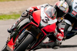
In another report from ISMRM 2023, you can find out about how MRI was used at the Motorcycle Grand Prix World Championship held in Valencia, Spain, in November 2022.
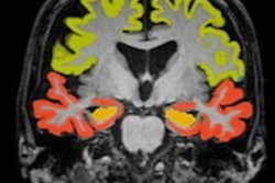
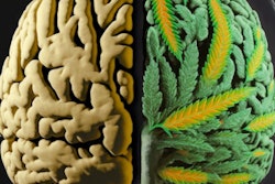
Cannabis (or marijuana) is one of the most widely used illicit drugs worldwide, and by 21 years of age, 80% of New Zealanders will have tried it at least once, with 10% going on to develop a pattern of heavy use or dependence, according to more research presented at ISMRM 2023. But what impact does this have on the brain? Learn more in our report.
Many of you will know Prof. Bettina Baessler, who was voted Most Effective Radiology Educator in the 2022 EuroMinnies awards. She gave a keynote talk about MRI radiomics on 3 June in Toronto, and it was well received.
An Italian investigation into how to cut MRI contrast residuals in hospital wastewater also caught our attention at ISMRM 2023. You can find out more in this article written by Kate Madden Yee, one of our three editors onsite.
In this letter, we've highlighted just a few of the many reports posted in the MRI Community over recent weeks. Please scroll through the full list below and contact me if you have ideas for future coverage.
Also, for more live reports from ISMRM, please check out the ISMRM Radcast on AuntMinnie.com.


![Overview of the study design. (A) The fully automated deep learning framework was developed to estimate body composition (BC) (defined as subcutaneous adipose tissue [SAT] in liters; visceral adipose tissue [VAT] in liters; skeletal muscle [SM] in liters; SM fat fraction [SMFF] as a percentage; and intramuscular adipose tissue [IMAT] in deciliters) from MRI. The fully automated framework comprised one model (model 1) to quantify different BC measures (SAT, VAT, SM, SMFF, and IMAT) as three-dimensional (3D) measures from whole-body MRI scans. The second model (model 2) was trained to identify standardized anatomic landmarks along the craniocaudal body axis (z coordinate field), which allowed for subdividing the whole-body measures into different subregions typically examined on clinical routine MRI scans (chest, abdomen, and pelvis). (B) BC was quantified from whole-body MRI in over 66,000 individuals from two large population-based cohort studies, the UK Biobank (UKB) (36,317 individuals) and the German National Cohort (NAKO) (30,291 individuals). Bar graphs show age distribution by sex and cohort. BMI = body mass index. (C) After the performance assessment of the fully automated framework, the change in BC measures, distributions, and profiles across age decades were investigated. Age-, sex-, and height-adjusted body composition reference curves were calculated and made publicly available in a web-based z-score calculator (https://circ-ml.github.io).](https://img.auntminnieeurope.com/mindful/smg/workspaces/default/uploads/2026/05/body-comp.XgAjTfPj1W.jpg?auto=format%2Ccompress&fit=crop&h=100&q=70&w=100)


![Overview of the study design. (A) The fully automated deep learning framework was developed to estimate body composition (BC) (defined as subcutaneous adipose tissue [SAT] in liters; visceral adipose tissue [VAT] in liters; skeletal muscle [SM] in liters; SM fat fraction [SMFF] as a percentage; and intramuscular adipose tissue [IMAT] in deciliters) from MRI. The fully automated framework comprised one model (model 1) to quantify different BC measures (SAT, VAT, SM, SMFF, and IMAT) as three-dimensional (3D) measures from whole-body MRI scans. The second model (model 2) was trained to identify standardized anatomic landmarks along the craniocaudal body axis (z coordinate field), which allowed for subdividing the whole-body measures into different subregions typically examined on clinical routine MRI scans (chest, abdomen, and pelvis). (B) BC was quantified from whole-body MRI in over 66,000 individuals from two large population-based cohort studies, the UK Biobank (UKB) (36,317 individuals) and the German National Cohort (NAKO) (30,291 individuals). Bar graphs show age distribution by sex and cohort. BMI = body mass index. (C) After the performance assessment of the fully automated framework, the change in BC measures, distributions, and profiles across age decades were investigated. Age-, sex-, and height-adjusted body composition reference curves were calculated and made publicly available in a web-based z-score calculator (https://circ-ml.github.io).](https://img.auntminnieeurope.com/mindful/smg/workspaces/default/uploads/2026/05/body-comp.XgAjTfPj1W.jpg?auto=format%2Ccompress&fit=crop&h=112&q=70&w=112)


