Dear MRI Insider,
Direct comparisons of images acquired on 7-tesla and 3-tesla MR systems are relatively scarce, so a new study by researchers from Lund University Hospital in Sweden was always going to attract considerable interest.
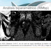
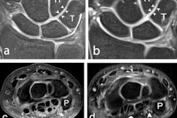
Injuries to the wrist are difficult to assess noninvasively, due to the wrist's small size and tricky anatomical structures such as intercarpal ligaments, the triangular fibrocartilage complex, and articular cartilage. However, the group found that 7-tesla MRI improves anatomical visualization and image quality over 3-tesla imaging, and this could lead to better detection and management of pathologies.
The early days of MRI continue to arouse controversy and divide opinion. Prof. Dr. Peter Rinck has written extensively about the topic for many years, and in a new column to mark the modality's 50th anniversary, he recalls a meeting that proved pivotal in the development of clinical MRI. Don't miss his fascinating article posted today.
Does MRI have a role to play in suspected cases of sepsis? Maybe, according to German expert Prof. Dr. Sebastian Ley. MRI can be helpful in certain situations, such as when it's necessary to reliably detect cerebral infections or spondylodiscitis. Find out more in this insightful interview.
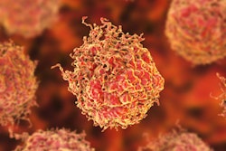
Meanwhile, the findings of an important prostate study were published by Lancet Oncology earlier this month. Scientists from the Karolinska Institute in Stockholm concluded that using a new type of blood test to screen for prostate cancer can reduce unnecessary MRI scans by 36% and avoid overdiagnosis.
The Paralympics get underway in Tokyo on 24 August, and MRI is likely to be used extensively at the event. The presence of metallic implants and other devices in these athletes looks set to represent a particular challenge, noted Dr. Yukihisa Saida in our most recent article about the Games.
This letter features only a few of the many reports posted in the MRI Community over the past few weeks. Please scroll through the full list below, and feel free to contact me if you have ideas for future coverage.


![Overview of the study design. (A) The fully automated deep learning framework was developed to estimate body composition (BC) (defined as subcutaneous adipose tissue [SAT] in liters; visceral adipose tissue [VAT] in liters; skeletal muscle [SM] in liters; SM fat fraction [SMFF] as a percentage; and intramuscular adipose tissue [IMAT] in deciliters) from MRI. The fully automated framework comprised one model (model 1) to quantify different BC measures (SAT, VAT, SM, SMFF, and IMAT) as three-dimensional (3D) measures from whole-body MRI scans. The second model (model 2) was trained to identify standardized anatomic landmarks along the craniocaudal body axis (z coordinate field), which allowed for subdividing the whole-body measures into different subregions typically examined on clinical routine MRI scans (chest, abdomen, and pelvis). (B) BC was quantified from whole-body MRI in over 66,000 individuals from two large population-based cohort studies, the UK Biobank (UKB) (36,317 individuals) and the German National Cohort (NAKO) (30,291 individuals). Bar graphs show age distribution by sex and cohort. BMI = body mass index. (C) After the performance assessment of the fully automated framework, the change in BC measures, distributions, and profiles across age decades were investigated. Age-, sex-, and height-adjusted body composition reference curves were calculated and made publicly available in a web-based z-score calculator (https://circ-ml.github.io).](https://img.auntminnieeurope.com/mindful/smg/workspaces/default/uploads/2026/05/body-comp.XgAjTfPj1W.jpg?auto=format%2Ccompress&fit=crop&h=100&q=70&w=100)


![Overview of the study design. (A) The fully automated deep learning framework was developed to estimate body composition (BC) (defined as subcutaneous adipose tissue [SAT] in liters; visceral adipose tissue [VAT] in liters; skeletal muscle [SM] in liters; SM fat fraction [SMFF] as a percentage; and intramuscular adipose tissue [IMAT] in deciliters) from MRI. The fully automated framework comprised one model (model 1) to quantify different BC measures (SAT, VAT, SM, SMFF, and IMAT) as three-dimensional (3D) measures from whole-body MRI scans. The second model (model 2) was trained to identify standardized anatomic landmarks along the craniocaudal body axis (z coordinate field), which allowed for subdividing the whole-body measures into different subregions typically examined on clinical routine MRI scans (chest, abdomen, and pelvis). (B) BC was quantified from whole-body MRI in over 66,000 individuals from two large population-based cohort studies, the UK Biobank (UKB) (36,317 individuals) and the German National Cohort (NAKO) (30,291 individuals). Bar graphs show age distribution by sex and cohort. BMI = body mass index. (C) After the performance assessment of the fully automated framework, the change in BC measures, distributions, and profiles across age decades were investigated. Age-, sex-, and height-adjusted body composition reference curves were calculated and made publicly available in a web-based z-score calculator (https://circ-ml.github.io).](https://img.auntminnieeurope.com/mindful/smg/workspaces/default/uploads/2026/05/body-comp.XgAjTfPj1W.jpg?auto=format%2Ccompress&fit=crop&h=112&q=70&w=112)


