Dear AuntMinnieEurope Member,
Estimating the risk of CT-induced cancers is notoriously difficult, but given the importance of the topic, it’s no surprise that researchers continue to tackle this arduous task.
The latest group to try has based its risk model on a multicenter sample of CT exams from the University of California, San Francisco’s International CT Dose Registry. From these scans, the team determined organ-specific radiation doses by patient age, sex, and CT category, then scaled the data to the U.S. population based on the number of exams in 2023. Check out the full story.
Sweden has a particularly rich history and strong track record in breast cancer screening, so the new cost-effectiveness study by the well-respected Prof. Sophia Zackrisson and her colleagues is bound to generate considerable interest.
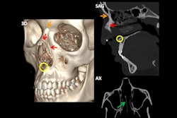
Spanish radiologists showed conclusively at ECR 2025 that imaging has a central role to play in challenging cases of facial trauma. First, presenters from San Sebastián won a magna cum laude award for their work in this area, and they’ve shared their experiences and clinical images with us.
A second Spanish team gave an impressive presentation at the Vienna congress about orbital findings that tend to be overlooked. It received a cum laude award for the exhibit.
Finally, a major industry story broke this week. RadNet has acquired AI software developer iCAD for around $103 million (€90.5 million), and it plans to integrate iCAD into its wholly owned AI subsidiary DeepHealth. iCAD's breast cancer risk assessment software has received the CE Mark, so this deal looks set to have an impact in Europe.
Philip Ward
Editor in Chief
AuntMinnieEurope.com