Dear AuntMinnieEurope Member,
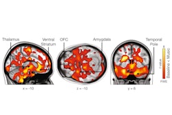
The influential French neuroscientist, radiologist, and pianist Prof. Denis Le Bihan has long had a keen interest in the benefits of music exposure on mental performance and cognitive development (see our article from 3 October 2022). Now a group from Turku, Finland, has built on his research and provided the first neuroimaging evidence that music affects the brain’s opioid receptor system.
This new finding is from an analysis in which 15 women underwent two PET scans, one before and then one while listening to their playlists, and it reveals new information on how the brain responds to sources of pleasure, according to the authors.
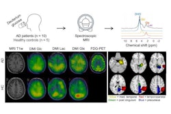
Another important study published this week comes from Denmark. The authors conducted a thorough investigation into the use of deuterium metabolic imaging in dementia. Their findings deserve a close look.
Let’s be honest, all of us love a good debate. Three well-known radiologists have expressed very different opinions about whether all trainees should be forced to undertake research. Each of them makes a very convincing case, as you can see in our special report.
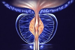
When it comes to imaging of the prostate, the Dutch have a formidable reputation, so new research from Nijmegen about image quality in prostate MRI is bound to generate plenty of interest. Check it out for yourself.
Finally, we bring you a report from outer space. The commander of the Fram2 mission has posted on social media what is thought to be the first x-ray image taken in space. Intriguingly, the image appears to be a nod to the first x-ray taken by Wilhelm Roentgen of his wife Anna Bertha Ludwig's hand on 22 December 1895.
Philip Ward
Editor in Chief
AuntMinnieEurope.com