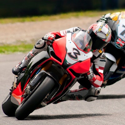
TORONTO - Portable MRI is proving to be a valuable tool at major sporting events -- including the Motorcycle Grand Prix (MotoGP) World Championship held in Valencia, Spain, in November of 2022.
The findings highlight how portable MRI is making the modality more versatile, noted an International Society for Magnetic Resonance in Medicine (ISMRM) presentation by doctoral candidate Teresa Guallart-Naval of the Universitat Politècnica de València in Spain.
"[Our] work demonstrates that portable MRI machines can aid the diagnostic capabilities of medical staff in sporting events and competitions," Guallart-Naval and colleagues told attendees at this week's annual meeting.
Low-field, portable MRI features permanent or resistive, rather than superconducting, magnets, the team noted, which allow for a lightweight design. This type of portable MRI technology may lend itself to a range of applications, including the following:
- Bedside and point-of care exams
- Outpatient services
- Emergency rooms and intensive care units
- Out-of-hospital exams for residential and hospice care
- Small clinics and rural areas
- Penitentiaries
- Sports clubs and school facilities
- Field hospitals and military camps
- Sporting events
The portable system was transported in a small truck, installed in the main surgery room of the race's medical facilities, and up and running 30 minutes later. The scanner weighs less than 250 kg -- including its electronics and a wheeled mounting platform -- and runs from a wall outlet.
The researchers used the device to scan 15 race participants over the four days of the race; these participants included racers, track marshals, medical staff, safety car drivers, helicopter pilots, event and race control staff, and engineers. The team performed 21 exams that included participants with injuries and healthy controls. Of these, five were of wrists (four healthy and one injured); 10 were of knees (four healthy and six injured); and six were of ankles (three healthy and three injured). Eight of these injuries had been diagnosed before the event.
3D-STIR MRI imaging confirmed the following:
- Bright volume between the scaphoid, trapezium, and trapezoid bones in the injured wrist
- Bone marrow edema and gonarthrosis in two of the injured knees
- An enlarged bony section of the heel near the Achilles tendon in two of the injured ankles and a bone fracture in one
"Out of eight low-field acquisitions on previously diagnosed lesions, only one was not detected by the circuit experts that evaluated our images," the group concluded. "This corresponds to a meniscus fracture, which may require higher spatial resolution than we currently achieve. Furthermore, we were able to detect a synovitis in the wrist which would have otherwise gone unnoticed to the MotoGP medical staff. Finally, no anomalies were detected in the scans performed on reportedly healthy volunteers."


![Examples of ultrasound findings and techniques. (A) Images in a 39-year-old male patient with a mass in the left thigh. The mass is heterogeneous on the B-mode US image (compared with the patient in D) and showed increased microvascularity (superb microvascular imaging [SMI]) and shear-wave elastography (SWE) values. Undifferentiated pleomorphic sarcoma was diagnosed at biopsy (with pleomorphic rhabdomyosarcoma in surgical specimen). (B) Images in an 18-year-old male patient with a mass in the left leg. The mass is hypoechoic on the B-mode image, with no other findings suggestive of malignancy. The lesion is in contact with the cortex of the tibia, which is slightly irregular. CT revealed a doubtful anteromedial tibial erosion. The microvascular study demonstrated high vascularization, suggestive of malignancy. Periosteal Ewing sarcoma was diagnosed with both histologic and immunohistochemical confirmation. (C) Images in a 69-year-old female patient with a lump growing on the outside of the left leg. Multiple SWE examinations were performed (please note the high values obtained in the measurements, whereas the color map highlights the stiffness relative to adjacent tissues). SMI showed areas of increased vascularization to target for sampling. Undifferentiated spindle cell sarcoma was diagnosed at biopsy, with residual leiomyosarcoma in the surgical specimen after neoadjuvant therapy. (D) Images in a 56-year-old female patient with a mass in the right thigh. The mass is heterogeneous at both B-mode ultrasound (similar to patient A) and MRI (coronal T2-weighted spectral attenuated inversion recovery [SPAIR]; T1-weighted pre-contrast and postcontrast imaging), which even shows uptake after the administration of paramagnetic contrast material, which is traditionally suggestive of malignancy. Low values at SMI and elastography are suggestive of benignity. Spindle cell lipoma was diagnosed at biopsy, with atypical spindle cell lipomatous tumor in the surgical specimen.](https://img.auntminnieeurope.com/mindful/smg/workspaces/default/uploads/2026/08/images-radiol250278fig2.APCFLSvX6p.jpg?auto=format%2Ccompress&fit=crop&h=100&q=70&w=100)


![Examples of ultrasound findings and techniques. (A) Images in a 39-year-old male patient with a mass in the left thigh. The mass is heterogeneous on the B-mode US image (compared with the patient in D) and showed increased microvascularity (superb microvascular imaging [SMI]) and shear-wave elastography (SWE) values. Undifferentiated pleomorphic sarcoma was diagnosed at biopsy (with pleomorphic rhabdomyosarcoma in surgical specimen). (B) Images in an 18-year-old male patient with a mass in the left leg. The mass is hypoechoic on the B-mode image, with no other findings suggestive of malignancy. The lesion is in contact with the cortex of the tibia, which is slightly irregular. CT revealed a doubtful anteromedial tibial erosion. The microvascular study demonstrated high vascularization, suggestive of malignancy. Periosteal Ewing sarcoma was diagnosed with both histologic and immunohistochemical confirmation. (C) Images in a 69-year-old female patient with a lump growing on the outside of the left leg. Multiple SWE examinations were performed (please note the high values obtained in the measurements, whereas the color map highlights the stiffness relative to adjacent tissues). SMI showed areas of increased vascularization to target for sampling. Undifferentiated spindle cell sarcoma was diagnosed at biopsy, with residual leiomyosarcoma in the surgical specimen after neoadjuvant therapy. (D) Images in a 56-year-old female patient with a mass in the right thigh. The mass is heterogeneous at both B-mode ultrasound (similar to patient A) and MRI (coronal T2-weighted spectral attenuated inversion recovery [SPAIR]; T1-weighted pre-contrast and postcontrast imaging), which even shows uptake after the administration of paramagnetic contrast material, which is traditionally suggestive of malignancy. Low values at SMI and elastography are suggestive of benignity. Spindle cell lipoma was diagnosed at biopsy, with atypical spindle cell lipomatous tumor in the surgical specimen.](https://img.auntminnieeurope.com/mindful/smg/workspaces/default/uploads/2026/08/images-radiol250278fig2.APCFLSvX6p.jpg?auto=format%2Ccompress&fit=crop&h=112&q=70&w=112)