Dear AuntMinnieEurope Member,
A broad approach can sometimes be beneficial in molecular imaging. New research from one of the leading centers for cancer imaging in Europe has found that PET/CT and MRI are better together for predicting patient outcomes in cervical cancer.
A group from Leeds Teaching Hospitals NHS Trust in the U.K. reported that the combination was more accurate than either modality alone for assessing response after chemotherapy in cases of locally advanced cervical carcinoma. Prof. Andy Scarsbrook, a consultant radiologist and nuclear medicine physician at St James's University Hospital and professor of radiology at the University of Leeds, served as one of the co-authors on the award-winning study.
In other molecular imaging news, a research team from Turkey and the U.S. has shown that cancer patients who receive COVID-19 mRNA vaccines have higher rates of reactive lymph nodes than those who received whole-virus vaccines. They warned that vaccine-related axillary lymph node enlargement could lead to unwarranted interventions or a delay in cancer care.
Within healthcare, radiology is an outsized consumer of energy. And MRI is one of imaging's most energy-intensive modalities. Fortunately, radiology departments can reduce carbon emissions caused to MRI by more than a third with one simple step, according to a recent study we covered in our MRI Community.
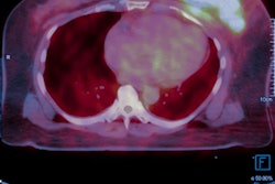
An artificial intelligence (AI) algorithm can detect cardiac amyloidosis on whole-body scintigraphy exams, potentially helping to identify thousands of undiagnosed cases, a group from France has revealed.
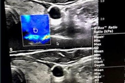
Meanwhile, researchers have found that an AI algorithm could offer significant value in breast ultrasound. A team from China reported that their model, which analyzes both grayscale and contrast-enhanced ultrasound, (CEUS) could perform comparably to ultrasound experts.
Speaking of ultrasound, the modality was also deemed to be a viable option for presurgical imaging of patients with primary hyperparathyroidism. In a prospective study, researchers from Denmark concluded that conventional ultrasonography and CEUS performed by an experienced parathyroid surgeon-sonographer was noninferior to scintigraphy.