Whole-body PET/CT scans could reduce radiation dose and significantly trim imaging time in melanoma patients, according to a study published on 7 December in the European Journal of Nuclear Medicine and Molecular Imaging.
A team led by Dr. Christos Sachpekidis at the German Cancer Research Center in Heidelberg, Germany, explored the capabilities of a new long axial field-of-view PET/CT scanner for imaging melanoma patients. Using a low dose of F-18 FDG radiotracer, the group reduced acquisition time almost by half, according to the study.
"Clinical introduction of ultra-extended field of view PET/CT scanners represents an evolution in molecular imaging with potentially major clinical and financial implications, while enabling, at the same time, a reduction of the associated radiation burden," the group wrote.
Recently, long axial field-of-view PET/CT systems, such as the Biograph Vision Quadra (Siemens Healthineers) used in this study and the uEXPLORER (United Imaging), have become available. Early experience shows these systems improve image quality, yet studies beyond research purposes are scarce, with initial clinical experiences just beginning to be reported.
In this study, Sachpekidis and colleagues aimed to determine an appropriate acquisition time for one of these new systems at their hospital in a group of melanoma patients after an F-18 FDG radiotracer injection of 2 MBq/kg rather than 3 MBq/kg as recommended by European guidelines.
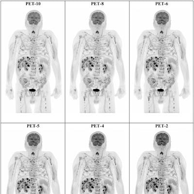
The investigators enrolled 49 melanoma patients, all of whom underwent whole-body PET/CT from the top of the head through the feet. images of standard 10-minute acquisition times were reconstructed and further split into eight-minute, six-minute, five-minute, four-minute, and two-minute durations (PET-10, -8, -6, -5, -4, and -2). The researchers then compared the different reconstructed scan times.
Example of maximum intensity projection (MIP) F-18 FDG-PET images of a 74-year-old man with stage IV cutaneous melanoma planned for treatment with immune checkpoint inhibitors. Presented are the PET-10, PET-8, PET-6, PET-5, PET-4, and PET-2 acquisitions. All acquisitions clearly demonstrate disseminated F-18 FDG-avid metastatic disease. Image and caption courtesy of the European Journal of Nuclear Medicine and Molecular Imaging.A total of 42 PET-10 scans showing a total of 119 tumor lesions were evaluated. The results of the visual analysis showed that PET-8 scans revealed 116 tumor lesions, PET-6 revealed 113, PET-5 revealed 107, PET-4 revealed 99, and PET-2 revealed 88 of 199 tumor lesions.
"The results of the visual analysis of our study show that this increased sensitivity of the new scanner may allow for a reasonable decrease in acquisition time by five minutes (i.e., from 10- to 5-min), even when applying low dose examination protocols," the researchers reported.
In standard clinical practice, acquiring high-quality PET/CT images requires either increasing the radiotracer injection dose or prolonging the scan time, which carry radiation exposure and undesirable logistical consequences, according to the authors. Whole-body low-dose long axial field-of-view PET/CT imaging may help overcome these limitations, they wrote.
"This may have significant implications for the diagnostic work-up of patients with melanoma, given the need for true whole-body imaging in this type of cancer," Sachpekidis and colleagues concluded.


![Examples of ultrasound findings and techniques. (A) Images in a 39-year-old male patient with a mass in the left thigh. The mass is heterogeneous on the B-mode US image (compared with the patient in D) and showed increased microvascularity (superb microvascular imaging [SMI]) and shear-wave elastography (SWE) values. Undifferentiated pleomorphic sarcoma was diagnosed at biopsy (with pleomorphic rhabdomyosarcoma in surgical specimen). (B) Images in an 18-year-old male patient with a mass in the left leg. The mass is hypoechoic on the B-mode image, with no other findings suggestive of malignancy. The lesion is in contact with the cortex of the tibia, which is slightly irregular. CT revealed a doubtful anteromedial tibial erosion. The microvascular study demonstrated high vascularization, suggestive of malignancy. Periosteal Ewing sarcoma was diagnosed with both histologic and immunohistochemical confirmation. (C) Images in a 69-year-old female patient with a lump growing on the outside of the left leg. Multiple SWE examinations were performed (please note the high values obtained in the measurements, whereas the color map highlights the stiffness relative to adjacent tissues). SMI showed areas of increased vascularization to target for sampling. Undifferentiated spindle cell sarcoma was diagnosed at biopsy, with residual leiomyosarcoma in the surgical specimen after neoadjuvant therapy. (D) Images in a 56-year-old female patient with a mass in the right thigh. The mass is heterogeneous at both B-mode ultrasound (similar to patient A) and MRI (coronal T2-weighted spectral attenuated inversion recovery [SPAIR]; T1-weighted pre-contrast and postcontrast imaging), which even shows uptake after the administration of paramagnetic contrast material, which is traditionally suggestive of malignancy. Low values at SMI and elastography are suggestive of benignity. Spindle cell lipoma was diagnosed at biopsy, with atypical spindle cell lipomatous tumor in the surgical specimen.](https://img.auntminnieeurope.com/mindful/smg/workspaces/default/uploads/2026/08/images-radiol250278fig2.APCFLSvX6p.jpg?auto=format%2Ccompress&fit=crop&h=100&q=70&w=100)


![Examples of ultrasound findings and techniques. (A) Images in a 39-year-old male patient with a mass in the left thigh. The mass is heterogeneous on the B-mode US image (compared with the patient in D) and showed increased microvascularity (superb microvascular imaging [SMI]) and shear-wave elastography (SWE) values. Undifferentiated pleomorphic sarcoma was diagnosed at biopsy (with pleomorphic rhabdomyosarcoma in surgical specimen). (B) Images in an 18-year-old male patient with a mass in the left leg. The mass is hypoechoic on the B-mode image, with no other findings suggestive of malignancy. The lesion is in contact with the cortex of the tibia, which is slightly irregular. CT revealed a doubtful anteromedial tibial erosion. The microvascular study demonstrated high vascularization, suggestive of malignancy. Periosteal Ewing sarcoma was diagnosed with both histologic and immunohistochemical confirmation. (C) Images in a 69-year-old female patient with a lump growing on the outside of the left leg. Multiple SWE examinations were performed (please note the high values obtained in the measurements, whereas the color map highlights the stiffness relative to adjacent tissues). SMI showed areas of increased vascularization to target for sampling. Undifferentiated spindle cell sarcoma was diagnosed at biopsy, with residual leiomyosarcoma in the surgical specimen after neoadjuvant therapy. (D) Images in a 56-year-old female patient with a mass in the right thigh. The mass is heterogeneous at both B-mode ultrasound (similar to patient A) and MRI (coronal T2-weighted spectral attenuated inversion recovery [SPAIR]; T1-weighted pre-contrast and postcontrast imaging), which even shows uptake after the administration of paramagnetic contrast material, which is traditionally suggestive of malignancy. Low values at SMI and elastography are suggestive of benignity. Spindle cell lipoma was diagnosed at biopsy, with atypical spindle cell lipomatous tumor in the surgical specimen.](https://img.auntminnieeurope.com/mindful/smg/workspaces/default/uploads/2026/08/images-radiol250278fig2.APCFLSvX6p.jpg?auto=format%2Ccompress&fit=crop&h=112&q=70&w=112)