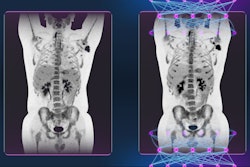
A multidisciplinary team of U.K. experts has produced a comprehensive guide to help determine when the clinical use of PET/CT is justified. The 86-page document can be downloaded free of charge.
"These guidelines mark the 30th anniversary of clinical PET in the U.K.," explained lead author Prof. Sabina Dizdarevic, chair of the Intercollegiate Standing Committee on Nuclear Medicine (ICSCNM). "PET-CT has become a key multimodality molecular imaging technique in the assessment of a wide range of medical conditions."
Now in its fourth edition, the 2022 document provides updated indications with key references underpinning the use of FDG and non-FDG PET/CT tracers in malignant and nonmalignant diseases in clinical practice. It was compiled by a team of nuclear medicine physicians, radionuclide radiologists, and oncologists, noted Dizdarevic, who is the principal lead consultant in imaging and nuclear medicine at the Brighton and Sussex University Hospitals NHS Trust and PET/CT lead at the Clinical Imaging Science Centre.
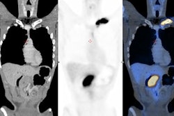
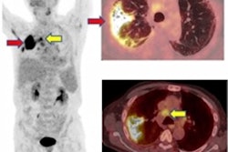
F-18 FDG PET/CT in the assessment of an infected cardiac pacemaker. (All figures courtesy of RCR)The guidelines comprise an up-to-date summary of relevant indications for the use of PET/CT, where there is good evidence that patients will benefit from improved disease assessment, resulting in altered management and improved outcomes, according to the authors.
The indications are divided into oncological and nononcological applications followed by body area/system. This list is not exhaustive, and there are cases where PET/CT may be helpful in patients who have equivocal or definite abnormalities on other imaging where PET/CT may alter the management strategy if found to be "positive" or "negative"; for example, radical or high-risk surgery, they added.
PET/CT would be appropriate in such patients at the discretion of the local Administration of Radioactive Substances Advisory Committee license holder as a problem-solving tool when other imaging modalities have been inconclusive.
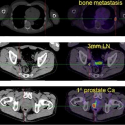
Gallium-68 (Ga-68)-labeled prostate-specific membrane antigen ligand PET/CT in prostate cancer.The new document supersedes the previous "Evidence-Based Indications for the Use of PET-CT in the United Kingdom" guidelines published by the Royal College of Radiologists (RCR) in 2016. The document will be updated at regular intervals.
The 2022 report was prepared for the ICSCNM, the RCR, and the Royal College of Physicians by Dizdarevic, Prof. Andy Scarsbrook from Leeds, Prof. Sally Barrington from King's College London, and other specialists.
You can download it free of charge from the RCR website.


![Examples of ultrasound findings and techniques. (A) Images in a 39-year-old male patient with a mass in the left thigh. The mass is heterogeneous on the B-mode US image (compared with the patient in D) and showed increased microvascularity (superb microvascular imaging [SMI]) and shear-wave elastography (SWE) values. Undifferentiated pleomorphic sarcoma was diagnosed at biopsy (with pleomorphic rhabdomyosarcoma in surgical specimen). (B) Images in an 18-year-old male patient with a mass in the left leg. The mass is hypoechoic on the B-mode image, with no other findings suggestive of malignancy. The lesion is in contact with the cortex of the tibia, which is slightly irregular. CT revealed a doubtful anteromedial tibial erosion. The microvascular study demonstrated high vascularization, suggestive of malignancy. Periosteal Ewing sarcoma was diagnosed with both histologic and immunohistochemical confirmation. (C) Images in a 69-year-old female patient with a lump growing on the outside of the left leg. Multiple SWE examinations were performed (please note the high values obtained in the measurements, whereas the color map highlights the stiffness relative to adjacent tissues). SMI showed areas of increased vascularization to target for sampling. Undifferentiated spindle cell sarcoma was diagnosed at biopsy, with residual leiomyosarcoma in the surgical specimen after neoadjuvant therapy. (D) Images in a 56-year-old female patient with a mass in the right thigh. The mass is heterogeneous at both B-mode ultrasound (similar to patient A) and MRI (coronal T2-weighted spectral attenuated inversion recovery [SPAIR]; T1-weighted pre-contrast and postcontrast imaging), which even shows uptake after the administration of paramagnetic contrast material, which is traditionally suggestive of malignancy. Low values at SMI and elastography are suggestive of benignity. Spindle cell lipoma was diagnosed at biopsy, with atypical spindle cell lipomatous tumor in the surgical specimen.](https://img.auntminnieeurope.com/mindful/smg/workspaces/default/uploads/2026/08/images-radiol250278fig2.APCFLSvX6p.jpg?auto=format%2Ccompress&fit=crop&h=100&q=70&w=100)


![Examples of ultrasound findings and techniques. (A) Images in a 39-year-old male patient with a mass in the left thigh. The mass is heterogeneous on the B-mode US image (compared with the patient in D) and showed increased microvascularity (superb microvascular imaging [SMI]) and shear-wave elastography (SWE) values. Undifferentiated pleomorphic sarcoma was diagnosed at biopsy (with pleomorphic rhabdomyosarcoma in surgical specimen). (B) Images in an 18-year-old male patient with a mass in the left leg. The mass is hypoechoic on the B-mode image, with no other findings suggestive of malignancy. The lesion is in contact with the cortex of the tibia, which is slightly irregular. CT revealed a doubtful anteromedial tibial erosion. The microvascular study demonstrated high vascularization, suggestive of malignancy. Periosteal Ewing sarcoma was diagnosed with both histologic and immunohistochemical confirmation. (C) Images in a 69-year-old female patient with a lump growing on the outside of the left leg. Multiple SWE examinations were performed (please note the high values obtained in the measurements, whereas the color map highlights the stiffness relative to adjacent tissues). SMI showed areas of increased vascularization to target for sampling. Undifferentiated spindle cell sarcoma was diagnosed at biopsy, with residual leiomyosarcoma in the surgical specimen after neoadjuvant therapy. (D) Images in a 56-year-old female patient with a mass in the right thigh. The mass is heterogeneous at both B-mode ultrasound (similar to patient A) and MRI (coronal T2-weighted spectral attenuated inversion recovery [SPAIR]; T1-weighted pre-contrast and postcontrast imaging), which even shows uptake after the administration of paramagnetic contrast material, which is traditionally suggestive of malignancy. Low values at SMI and elastography are suggestive of benignity. Spindle cell lipoma was diagnosed at biopsy, with atypical spindle cell lipomatous tumor in the surgical specimen.](https://img.auntminnieeurope.com/mindful/smg/workspaces/default/uploads/2026/08/images-radiol250278fig2.APCFLSvX6p.jpg?auto=format%2Ccompress&fit=crop&h=112&q=70&w=112)