PET/CT is developing an important clinical role in the diagnosis and follow-up of patients with musculoskeletal (MSK) involvement from hematological and oncological diseases, Spanish researchers have reported.
"Radiologists must be familiar with these imaging studies to better understand the management of musculoskeletal lesions," noted Dr. Patricia García and Dr. Victor Rodríguez Laval, from the radiology department at La Princesa Hospital in Madrid.
They added that it is also essential to know about the role of PET/CT in the follow-up of hematological diseases – particularly lymphoma, plasmacytoma, and multiple myeloma – and oncological pathologies, namely primary tumors and metastases.
A decade of PET/CT studies
The authors reviewed the PET/CT studies from the last 10 years at their hospital. They presented their findings in a poster presentation at ECR 2022.
Epidural space is the compartment most often affected by hematological neoplasm and primary bone tumors, and multiple myeloma is the most common primary bone neoplasm and the most frequent primary bone tumor that may lead to spinal cord compression. In these cases, MRI is the gold standard for diagnosis and PET/CT plays an important role in staging or follow-up, depending on the neoplasm of origin, the researchers stated.
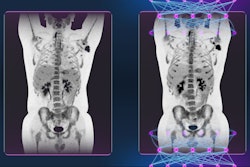
 A 57-year-old woman with monoclonal gammopathy of undetermined significance. Progression to multiple myeloma was suspected. PET/CT confirmed the diagnosis by showing pathological uptake of FDG in the humerus and vertebra. All figures courtesy of Dr. Patricia García, Dr. Victor Rodríguez Laval, et al, presented at ECR 2022.
In bone lesions, whole-body CT is a good initial examination for patients who have multiple myeloma. "If there are one or more 5-mm or larger lytic myeloma lesions, there is no need for the patient to undergo further investigation, with the exception of complication assessment, or treatment response evaluation, where the PET/CT plays an important role," they noted.
If a whole-body CT exam identifies no bone lesions, a follow-up investigation with whole-body MRI should be performed to exclude lesions and confirm the diagnosis of smoldering multiple myeloma, or to find lesions that are undetectable on whole-body CT to upgrade the diagnosis to multiple myeloma.
PET is useful to distinguish benign from malignant MSK tumors because low uptake suggests that a bone lesion is likely to be benign (false negatives in plasmacytoma and low-grade chondrosarcoma).
Chondrosarcomas usually have lower uptake than other sarcomas, but higher uptake than enchondromas. PET cannot distinguish between benign tumors and grade 1 chondrosarcomas. Grades 2 and 3 have higher glucose metabolism than low-grade cartilage tumors, according to the authors.
Soft-tissue lesions
In a known case of soft tissue lesion, PET is of some value in determining whether the lesion is benign or malignant and in grading malignant lesions. "Low uptake of FDG is of limited value in differentiating benign from malignant but high uptake usually indicates intermediate or high-grade malignancy. Radiographic correlation can often differentiate benign lesions with high uptake from sarcomas," they wrote.
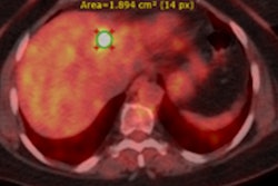
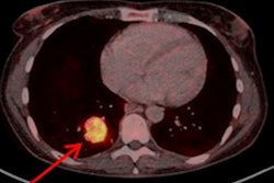
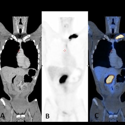
 A 26-year-old man with fever and night sweats. Coronal CT (A), PET (B), and fusion (C) images show intense uptake of FDG in the left clavicle. CT (D), PET (E), and fusion (F) images show intense uptake of FDG of a mass next to the pancreas. The biopsy was compatible with Burkitt lymphoma (non-Hodgkin's lymphoma).
Therefore PET is valuable in soft tissue malignancies for staging, guiding biopsy, detecting recurrence, therapy response, and tumor grading, and it is a standard modality for staging both Hodgkin's disease and non-Hodgkin's lymphoma. In Hodgkin's disease, PET can be of value in any stage, but it is most useful in stage 1 and 2 disease, where a change in the stage will alter disease management, the researchers pointed out.
"PET is particularly useful in later stages of the disease, when response to chemotherapy is substantially lower than in early stages," they noted. "PET is also useful as a prognostic indicator after one or a few cycles or at mid treatment, during first line chemotherapy of Hodgkin's disease and non-Hodgkin's lymphoma; lack of response suggests that the treatment regimen should be changed."
Follow-up research
The group is now conducting a follow-up study about the strengths and weaknesses of 18F-FDG-PET/CT in musculoskeletal involvement in oncological diseases, and it has been accepted by RSNA 2022 as a poster presentation, García told AuntMinnieEurope.com.
"We are going to illustrate the spectrum of hematological diseases and oncological pathologies with musculoskeletal involvement in FDG-PET/CT, including primary musculoskeletal tumors (osteosarcoma, Ewing, etc.), multiple myeloma and lymphoma, and metastasis and complications," García said. "We will discuss the importance of FDG-PET/CT in each pathology and its limitations."
Most of the PET/CT reports were carried out jointly with their nuclear medicine colleagues, Dr. V. Castillo and Dr. I. Hernández, who are both consultants and who contributed to the research, she added. The other coauthors of this research were Dr. M.C. Iniesta González, Dr. M.J. Moreno Casado, and Dr. N. Gómez Leon.
You can view the full ECR exhibit on the EPOS section of the European Society of Radiology website.