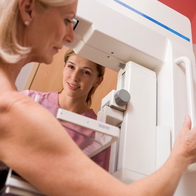
The benefits of mammography outweigh the risks of overdiagnosis -- especially since the overdiagnosis rate is low, a study published on 18 July in Cancer Medicine has found.
A study led by Dr. Oleg Blyuss from Queen Mary University of London included more than 57,000 women who participated in the U.K.'s National Health Service (NHS) Breast Screening program. It revealed that just three per 1,000 women had an overdiagnosis, corresponding to an estimated 3.7% overdiagnosis rate for screen-detected cancers.
"Our results showed little if any overdiagnosis, and it is reasonable to conclude that [the program] is associated with at worst modest overdiagnosis of breast cancer," Blyuss and colleagues wrote.
While breast cancer incidence continues to rise, mortality is on the decline, with researchers lauding improved treatment availability and earlier diagnosis by mammography screening. But overdiagnosis of the disease has been a topic of debate among breast imagers, and previous studies suggest there is a large variation as to how many women are overdiagnosed.
The variation is partly due to employing different methods used to calculate overdiagnosis, the researchers explained. Such methods include whether DCIS cancers are included, whether there is a sufficient follow-up period after stopping screening, the method used to adjust for lead time, and which denominator is used.
Blyuss and colleagues sought to use case-control data to explore the effect of attendance at screening on breast cancer incidence. They also wanted to use national incidence data to estimate the absolute level of overdiagnosis.
The team looked at data from 57,493 women in the cases cohort who were enrolled in the NHS program, which offers women mammography screening every three years. A total of 105,653 controls were also included. The women in the cases cohort had an age range of 47 to 89 years and were diagnosed with primary breast cancer, as well as invasive or ductal carcinoma in situ (DCIS) between 2010 and 2011.
The researchers found that the estimated number of overdiagnosed breast cancer cases in women attending all screens in the program was 679.3 per 100,000 without adjustment for self-selection bias. With that adjustment, however, the number decreased to 261.2 per 100,000 cases. This corresponded to an estimated 9.5% of screen-detected cancers overdiagnosed without adjustment and 3.7% with adjustment for self-selection.
The authors wrote that the absolute probability of having an overdiagnosed breast cancer is a more useful measure for women deciding whether to accept breast cancer screening offers.
"Even our more pessimistic estimate of overdiagnosis was lower than suggested in the past," they added.
The investigators did note that their findings focus on a program that offers mammography every three years from ages 50 to 70. They added that a more intensive program, such as annual mammography seen in the U.S., as well as screening with higher age limits, could result in higher overdiagnosis numbers.


![Examples of ultrasound findings and techniques. (A) Images in a 39-year-old male patient with a mass in the left thigh. The mass is heterogeneous on the B-mode US image (compared with the patient in D) and showed increased microvascularity (superb microvascular imaging [SMI]) and shear-wave elastography (SWE) values. Undifferentiated pleomorphic sarcoma was diagnosed at biopsy (with pleomorphic rhabdomyosarcoma in surgical specimen). (B) Images in an 18-year-old male patient with a mass in the left leg. The mass is hypoechoic on the B-mode image, with no other findings suggestive of malignancy. The lesion is in contact with the cortex of the tibia, which is slightly irregular. CT revealed a doubtful anteromedial tibial erosion. The microvascular study demonstrated high vascularization, suggestive of malignancy. Periosteal Ewing sarcoma was diagnosed with both histologic and immunohistochemical confirmation. (C) Images in a 69-year-old female patient with a lump growing on the outside of the left leg. Multiple SWE examinations were performed (please note the high values obtained in the measurements, whereas the color map highlights the stiffness relative to adjacent tissues). SMI showed areas of increased vascularization to target for sampling. Undifferentiated spindle cell sarcoma was diagnosed at biopsy, with residual leiomyosarcoma in the surgical specimen after neoadjuvant therapy. (D) Images in a 56-year-old female patient with a mass in the right thigh. The mass is heterogeneous at both B-mode ultrasound (similar to patient A) and MRI (coronal T2-weighted spectral attenuated inversion recovery [SPAIR]; T1-weighted pre-contrast and postcontrast imaging), which even shows uptake after the administration of paramagnetic contrast material, which is traditionally suggestive of malignancy. Low values at SMI and elastography are suggestive of benignity. Spindle cell lipoma was diagnosed at biopsy, with atypical spindle cell lipomatous tumor in the surgical specimen.](https://img.auntminnieeurope.com/mindful/smg/workspaces/default/uploads/2026/08/images-radiol250278fig2.APCFLSvX6p.jpg?auto=format%2Ccompress&fit=crop&h=100&q=70&w=100)


![Examples of ultrasound findings and techniques. (A) Images in a 39-year-old male patient with a mass in the left thigh. The mass is heterogeneous on the B-mode US image (compared with the patient in D) and showed increased microvascularity (superb microvascular imaging [SMI]) and shear-wave elastography (SWE) values. Undifferentiated pleomorphic sarcoma was diagnosed at biopsy (with pleomorphic rhabdomyosarcoma in surgical specimen). (B) Images in an 18-year-old male patient with a mass in the left leg. The mass is hypoechoic on the B-mode image, with no other findings suggestive of malignancy. The lesion is in contact with the cortex of the tibia, which is slightly irregular. CT revealed a doubtful anteromedial tibial erosion. The microvascular study demonstrated high vascularization, suggestive of malignancy. Periosteal Ewing sarcoma was diagnosed with both histologic and immunohistochemical confirmation. (C) Images in a 69-year-old female patient with a lump growing on the outside of the left leg. Multiple SWE examinations were performed (please note the high values obtained in the measurements, whereas the color map highlights the stiffness relative to adjacent tissues). SMI showed areas of increased vascularization to target for sampling. Undifferentiated spindle cell sarcoma was diagnosed at biopsy, with residual leiomyosarcoma in the surgical specimen after neoadjuvant therapy. (D) Images in a 56-year-old female patient with a mass in the right thigh. The mass is heterogeneous at both B-mode ultrasound (similar to patient A) and MRI (coronal T2-weighted spectral attenuated inversion recovery [SPAIR]; T1-weighted pre-contrast and postcontrast imaging), which even shows uptake after the administration of paramagnetic contrast material, which is traditionally suggestive of malignancy. Low values at SMI and elastography are suggestive of benignity. Spindle cell lipoma was diagnosed at biopsy, with atypical spindle cell lipomatous tumor in the surgical specimen.](https://img.auntminnieeurope.com/mindful/smg/workspaces/default/uploads/2026/08/images-radiol250278fig2.APCFLSvX6p.jpg?auto=format%2Ccompress&fit=crop&h=112&q=70&w=112)