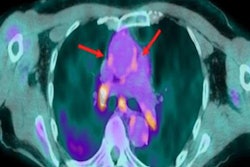
Contrast agent pooling on CT -- known as the CAP sign -- suggests patients are in danger of imminent cardiac arrest, according to a study published in BMC Emergency Medicine.
The findings may help clinicians intervene more quickly to prevent cardiac arrest in very sick patients, wrote a team led by Yu-Hsuan Lee, PhD, of Far Eastern Memorial Hospital in New Taipei City, Taiwan.
"The CAP sign can be considered as an alarm for imminent cardiac arrest and poor prognosis," the group wrote.
Cardiac arrest in hospitalized patients tends to be an unpredictable event, and clinicians have sought evidence-based ways to forecast it, the group noted. CAP identified on CT indicates blood leakage out of vessels and has been suggested as a precursor of sudden cardiac arrest in some studies, but clear evidence of the connection has not been established.
Therefore, Lee and colleagues sought to investigate the accuracy of the CAP sign on CT for predicting imminent cardiac arrest and association of the sign with patient survival.
The study included data from 128 patients who presented in the emergency room between January 2016 and December 2018, underwent contrast chest or abdominal CT and then experienced cardiac arrest. Reasons for the CT exam included such conditions as hypovolemic shock caused by trauma or internal bleeding, pulmonary embolism, heart attack, constrictive pericarditis, and bilateral pleural effusion.
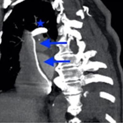
 Contrast agent pooling and layering over the inferior vena cava (arrow). B Contrast agent pooling over the right renal vein (arrow). C Retrograde pooling of the contrast agent over the dependent part of the hepatic veins and parenchyma (star). D Contrast agent layering over the superior vena cava (arrows) and pooling over the arch of the azygos vein (star). Images and caption courtesy of BMC Emergency Medicine through Creative Commons Attribution 4.0 International License.
The primary outcome of the study was whether cardiac arrest occurred within the hour of a positive CAP sign, with the secondary outcome patient survival and discharge.
Of the 128 patients, 11 (8.6%) had a positive CAP sign. A positive CAP sign had an accuracy of 85.9% for predicting cardiac arrest within an hour, the team found. The odds ratio for those with a positive CAP sign on CT having an episode of cardiac arrest at all was 7.35, and the odds ratio for within an hour was 12.8 (with 1 as reference).
| CAP sign on CT performance for predicting cardiac arrest | |
| Measure | Percentage |
| Accuracy | 85.9% |
| Negative predictive value | 88% |
| Positive predictive value | 64% |
| Specificity | 96% |
Of those patients with a positive CAP sign on CT, 63.7% experienced cardiac arrest within an hour; the remainder experienced it within eight hours, Lee's team reported. All of these patients died.
The CAP sign shows the effect of gravity, the group explained in the article posted on 6 May.
"The density of contrast agent is much higher than that of blood; thus, during normal physiological blood flow, specific gravity has little effect on the contrast agent dynamics," the team wrote. "In patients with cardiogenic shock or impending circulatory collapse, both the arterial and venous blood flow dramatically decreases, and the contrast agent tends to accumulate in the dependent parts of the venous system."
The CAP sign on CT offers an early warning for cardiac arrest for the very ill, according to the authors.
"From our findings, the CAP sign could be addressed as an imaging feature of circulatory failure, it could be used to predict imminent cardiac arrest and should be considered a warning sign for clinical physicians to provide in-time interventions for critically ill patients," they concluded.