More than half of people who contract COVID-19 pneumonia show evidence on chest CT of damage to their lungs even a year after symptom onset, according to a study published on 29 March in Radiology.
The findings underscore the importance of imaging follow-up in people who have apparently recovered from COVID-19, said study senior author Dr. Gerlig Widmann of Innsbruck Medical University in Austria in a statement released by the RSNA.
"Long-term follow-up, both clinical and radiological, is necessary to gather more information about the course and clinical role of persisting SARS-CoV-2 related chest CT abnormalities," he said.
The fact that pneumonia can damage the lungs is well known, but what is less understood is how COVID-19 pneumonia in particular affects the lungs long-term, said study co-author Dr. Anna Luger of the Medical University of Innsbruck in Austria in the statement.
"The observed chest CT abnormalities from our study are indicative of damaged lung tissue," she said. "However, it is currently unclear if they represent persistent scarring, and whether they regress over time or lead to pulmonary fibrosis.
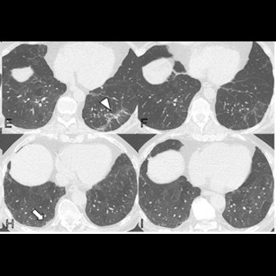
 Serial noncontrast axial chest CT exams of three study participants with prior COVID-19 pneumonia. Chest CT of a 44-year-old man (upper row, A-C) displayed extensive bilateral ground-glass opacities (GGO) and supleural reticulation during acute COVID-19 (A). At the two-month follow-up almost complete resolution of GGO with residual subpleural reticulation in the middle lobe was noted (B). These subpleural reticulations (arrow) persisted up to one year after onset (C). Chest CT of a 68-year-old-man (middle row, D-F) demonstrated patchy bilateral consolidations, a subpleural arcade-like sign, and pleural effusions during active infection (D). At the two-month follow-up, a substantial improvement of organizing pneumonia pattern was noted with GGO and subpleural reticulation including arcade-like sign (arrowhead) in the left lower lobe (E). At the one-year follow-up, further improvement was noticed. However, subtle reticulation and GGO could still be detected (F). Chest CT of a 79-year-old man (lower row, G-I) displayed bilateral consolidations and small areas of GGO while admitted to the intensive care unit (G). At the two-month follow-up, residual GGO and small subpleural microcystic changes (thick arrow) were noticed (H), which persisted up to one year after onset (I). Images and caption courtesy of the RSNA.
Luger's group conducted a study that included 91 patients evaluated with chest CT after contracting COVID-19 pneumonia at two, three, six, and 12 months after symptom onset. Of these, 49 (54%) showed abnormalities on chest CT at the one-year mark. CT exam findings were graded by lung lobe using a severity score that ranged from 0 (normal) to 25 (all lobes involved).
The group found the following:
- 54% of patients who contracted COVID-19 pneumonia had abnormalities on chest CT at one year post symptom onset.
- 51% of these patients with long-term COVID-19-related chest CT abnormalities were treated for the illness in a general hospital ward, while 45% were treated in the intensive care unit and 4% were treated on an outpatient basis only.
- 63% of patients with COVID-19 related abnormalities did not show further improvements after six months.
- Common abnormal findings were subtle subpleural reticulation, ground-glass opacities, or both (34%); 20% of the abnormal CT exams showed extensive ground-glass opacities, reticulations, bronchial dilation, or microcystic changes.
- Men older than 60 were particularly vulnerable to showing persistent lung abnormalities on CT at one year after symptom onset.
The study highlights the need to track individuals who have contracted COVID-19 pneumonia over time, according to the authors.
"Our results emphasize early and longitudinal monitoring of COVID-19 [patients]," they wrote. "[There] is still an urgent need for further studies focusing on histological and clinical correlations within the first three months after COVID-19 to identify [those] at risk for developing CT abnormalities and who would benefit from early tailored therapeutic concepts."