Dear AuntMinnieEurope Member,
If ever our editorial team doubted the hunger and desire for an awards scheme during a time when hospitals and clinics across Europe are under serious pressure, then you've given us an emphatic answer over the last few weeks -- you definitely want EuroMinnies 2022!
When we asked you for your nominations, you responded in style by sending us a record number of submissions and some intriguing choices. And now that we've released the names of the semifinal candidates, you've reacted enthusiastically on social media and via email.
My colleagues and I really appreciate your support and participation. Our expert panel comprising editorial advisory board members, past winners, and columnists is ready and waiting to cast their votes, and we'll reveal the finalists in January. Together, I'm sure we'll make this an awards scheme to remember and provide radiology with the recognition it deserves.
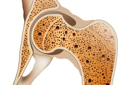
In other news, osteoporosis researchers from the world-famous Goethe University Frankfurt in Germany have found that quantitative analysis of bone mineral density on dual-energy CT has yielded impressive results. Don't miss our report from RSNA 2021.
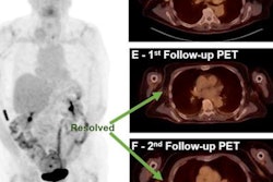
Austrian investigators have been keeping busy too. A team from Salzburg analyzed data from F-18 FDG-PET/CT scans in patients with metastatic melanoma before and after they began immunotherapy. They found that PET/CT scans performed a few months after patients began therapy predicted three- and five-year overall survival rates.
In our MRI Community, we have a report about Turkish scientists who tested artificial intelligence algorithms based on two convolutional neural networks. They found the models achieved high performance for both the detection of ischemic stroke and the classification of its vascular territorial type on diffusion-weighted MRI.
Last but not least, a Dutch group compared point shear-wave ultrasound elastography techniques on different systems to measure phantom elasticity in chronic liver disease patients who present with fibrosis.