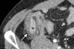
Appendicitis can be diagnosed with a low-dose CT protocol just as well as with CT at a standard radiation dose, according to a study published on 11 November in the British Journal of Surgery. Low-dose CT can also differentiate between cases that likely will require surgery.
The study's results mean that radiation exposure for patients with suspected appendicitis can be lessened -- good news, particularly since most appendicitis patients are younger, wrote a team led by Dr. Jussi Haijanen of Turku University Hospital in Finland.
"[The] radiation exposure associated with CT is an indisputable disadvantage, especially as the majority of patients with suspected acute appendicitis are young adults," the group wrote. "[As even] a small reduction in radiation dose reduces the risks of radiation in a large patient population, the principle of using as low a dose as reasonably achievable should be adhered to, with the aim of reducing radiation dose whenever possible."
There's increasing evidence that some cases of appendicitis can be treated in less invasive ways than surgery. But this makes the need for accurate diagnosis of the severity of the condition even more important, the authors noted. (Complicated acute appendicitis tends to be defined as disease that presents with perforation, tumor, gangrene, or abscess and requires surgery.)
"As emergency appendicectomy is no longer considered the only treatment alternative for patients with uncomplicated acute appendicitis, the emphasis on appendicitis diagnostics has shifted from solely assessing whether the patient has [the condition] or not towards differentiating between uncomplicated and complicated acute [disease]," they wrote.
Contrast-enhanced standard-dose CT is typically used to diagnose acute appendicitis, but Haijanen's team sought to explore whether using less radiation would prove just as effective. The study included 856 patients (median age, 37), of whom 454 (53%) underwent low-dose CT and 402 (47%) underwent a standard dose exam for suspected appendicitis between April 2017 and November 2018.
The investigators found that the overall accuracy of low-dose CT for identifying patients with and without acute appendicitis was essentially the same as the accuracy of standard-dose CT, at 98% and 98.5%, respectively. The accuracy for distinguishing between uncomplicated and complicated appendicitis was 90.3% for low-dose CT and 87.6% for standard dose. Mean radiation dose for the low-dose exams was 3 mSv, compared with 7 mSv for the standard dose exams.
The study findings demonstrate that CT radiation dose can be significantly decreased without negative effect on diagnostic accuracy, said lead author Dr. Paulina Salminen of the University of Turku in a statement released by the journal.
"These findings will hopefully encourage physicians to implement low-dose CT modalities at emergency departments for acute appendicitis imaging to avoid unnecessary radiation in this ... patient population," she said.