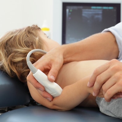
The British Institute of Radiology (BIR) has released a free podcast that focuses on training and best practice in musculoskeletal (MSK) ultrasound.
The podcast consists of a 37-minute interview with two leading U.K. experts: Dr. Ramy Mansour, clinical lead of MSK imaging at Oxford University Hospitals, and Dr. Danoob Dalili, consultant MSK radiologist from London. They were co-authors of the guidelines and recommendations paper, "Musculoskeletal ultrasound imaging standards in the UK: a British Society of Skeletal Radiologists position statement," published in April by the British Journal of Radiology.
 Dr. Danoob Dalili.
"The purpose [of this statement] was to review current practices affecting the delivery of MSK ultrasound and through that, provide recommendations for education, training, audit, clinical governance, reporting, and medico-legal issues," Dalili said. "It's important to emphasize that this statement is not restricting practice but essentially describing best practice models and campaigning for this to become a gold standard of care regardless of who sees the patient."
The podcast discusses the article, the results of a recent survey to gauge views on training opportunities in MSK radiology and ultrasound, and guidance for action from the position statement. It also focuses on future challenges, including how the experts see the field progressing in the future.
 Dr. Ramy Mansour.
"I expect that MSK ultrasound service and demand will continue to grow," said Mansour, who is MSK lead of the European Diploma in Radiology (EDiR) and the current British Society of Skeletal Radiology (BSSR) Royal College of Radiologists (RCR) Travelling Professor.
"I hope that this position statement will help standardize and improve the quality of the MSK ultrasound service that is provided for patients and encourage equal access to specialist MSK ultrasound service for all UK citizens. This can be achieved by standardized training, practices and competencies which are required to deliver a high-quality MSK ultrasound service in the U.K.," he noted.
In May 2021, Dalili, Mansour, and colleagues also published the findings of a survey on MSK training.

![Examples of ultrasound findings and techniques. (A) Images in a 39-year-old male patient with a mass in the left thigh. The mass is heterogeneous on the B-mode US image (compared with the patient in D) and showed increased microvascularity (superb microvascular imaging [SMI]) and shear-wave elastography (SWE) values. Undifferentiated pleomorphic sarcoma was diagnosed at biopsy (with pleomorphic rhabdomyosarcoma in surgical specimen). (B) Images in an 18-year-old male patient with a mass in the left leg. The mass is hypoechoic on the B-mode image, with no other findings suggestive of malignancy. The lesion is in contact with the cortex of the tibia, which is slightly irregular. CT revealed a doubtful anteromedial tibial erosion. The microvascular study demonstrated high vascularization, suggestive of malignancy. Periosteal Ewing sarcoma was diagnosed with both histologic and immunohistochemical confirmation. (C) Images in a 69-year-old female patient with a lump growing on the outside of the left leg. Multiple SWE examinations were performed (please note the high values obtained in the measurements, whereas the color map highlights the stiffness relative to adjacent tissues). SMI showed areas of increased vascularization to target for sampling. Undifferentiated spindle cell sarcoma was diagnosed at biopsy, with residual leiomyosarcoma in the surgical specimen after neoadjuvant therapy. (D) Images in a 56-year-old female patient with a mass in the right thigh. The mass is heterogeneous at both B-mode ultrasound (similar to patient A) and MRI (coronal T2-weighted spectral attenuated inversion recovery [SPAIR]; T1-weighted pre-contrast and postcontrast imaging), which even shows uptake after the administration of paramagnetic contrast material, which is traditionally suggestive of malignancy. Low values at SMI and elastography are suggestive of benignity. Spindle cell lipoma was diagnosed at biopsy, with atypical spindle cell lipomatous tumor in the surgical specimen.](https://img.auntminnieeurope.com/mindful/smg/workspaces/default/uploads/2026/08/images-radiol250278fig2.APCFLSvX6p.jpg?auto=format%2Ccompress&fit=crop&h=100&q=70&w=100)






![Examples of ultrasound findings and techniques. (A) Images in a 39-year-old male patient with a mass in the left thigh. The mass is heterogeneous on the B-mode US image (compared with the patient in D) and showed increased microvascularity (superb microvascular imaging [SMI]) and shear-wave elastography (SWE) values. Undifferentiated pleomorphic sarcoma was diagnosed at biopsy (with pleomorphic rhabdomyosarcoma in surgical specimen). (B) Images in an 18-year-old male patient with a mass in the left leg. The mass is hypoechoic on the B-mode image, with no other findings suggestive of malignancy. The lesion is in contact with the cortex of the tibia, which is slightly irregular. CT revealed a doubtful anteromedial tibial erosion. The microvascular study demonstrated high vascularization, suggestive of malignancy. Periosteal Ewing sarcoma was diagnosed with both histologic and immunohistochemical confirmation. (C) Images in a 69-year-old female patient with a lump growing on the outside of the left leg. Multiple SWE examinations were performed (please note the high values obtained in the measurements, whereas the color map highlights the stiffness relative to adjacent tissues). SMI showed areas of increased vascularization to target for sampling. Undifferentiated spindle cell sarcoma was diagnosed at biopsy, with residual leiomyosarcoma in the surgical specimen after neoadjuvant therapy. (D) Images in a 56-year-old female patient with a mass in the right thigh. The mass is heterogeneous at both B-mode ultrasound (similar to patient A) and MRI (coronal T2-weighted spectral attenuated inversion recovery [SPAIR]; T1-weighted pre-contrast and postcontrast imaging), which even shows uptake after the administration of paramagnetic contrast material, which is traditionally suggestive of malignancy. Low values at SMI and elastography are suggestive of benignity. Spindle cell lipoma was diagnosed at biopsy, with atypical spindle cell lipomatous tumor in the surgical specimen.](https://img.auntminnieeurope.com/mindful/smg/workspaces/default/uploads/2026/08/images-radiol250278fig2.APCFLSvX6p.jpg?auto=format%2Ccompress&dpr=2&fit=crop&h=167&q=70&w=250)


