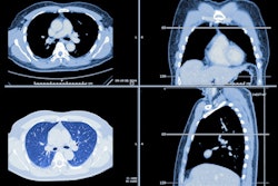
Using low-dose CT (LDCT) to screen for lung cancer in current and formerly heavy smokers offers more benefit than harm, according to the German Institute for Quality and Efficiency in Health Care (IQWiG). The final report commissioned by the Federal Joint Committee (G-BA) found LDCT can save a number of people from dying of lung cancer and, for some, prolong overall survival.
Lung cancer is usually discovered at a late stage so often only one-fifth to one-sixth of people affected are still alive within five years. Germany doesn't currently offer systematic screening for lung cancer, but LDCT could fill the void. The IQWiG investigated this possibility by analyzing eight randomized controlled trials with more than 90,000 people. The results show within about 10 years, LDCT can prevent about five out of every 1,000 people from dying of lung cancer.
However, LDCT doesn't show a statistical advantage for all-cause mortality -- perhaps because people saved from lung cancer death die from other tobacco-related diseases instead.
When it comes to a population-based screening program, two questions always come to mind: What is the misdiagnosis as well as overdiagnosis rate? The IQWiG found variable rates: one to 15 out of every 1,000 people underwent unnecessary surgery due to false-positive screening results. Similarly, the overdiagnosis risk varied: Out of every 1,000 people invited to screen, zero to 22 might be affected by overdiagnosis. The risk of overdiagnosis for people diagnosed with lung cancer during the screening phase ranged between 0% and 63%.
Given everything the institute found, the IQWiG concluded that there is a hint of a benefit in using LDCT for lung cancer screening, particularly for current and formerly heavy smokers versus no screening. However, screening should be supported by specific quality assurance measures such as diagnostic algorithm, types of devices, follow-up, etc. Additionally, more information will come to light in the next few years that could turn the tide for LDCT. Up to the end of 2024, a European study is investigating the safety of risk-based examination intervals and strategies for implementation: The "4-In The Lung Run" includes a total of 24,000 people.


![Examples of ultrasound findings and techniques. (A) Images in a 39-year-old male patient with a mass in the left thigh. The mass is heterogeneous on the B-mode US image (compared with the patient in D) and showed increased microvascularity (superb microvascular imaging [SMI]) and shear-wave elastography (SWE) values. Undifferentiated pleomorphic sarcoma was diagnosed at biopsy (with pleomorphic rhabdomyosarcoma in surgical specimen). (B) Images in an 18-year-old male patient with a mass in the left leg. The mass is hypoechoic on the B-mode image, with no other findings suggestive of malignancy. The lesion is in contact with the cortex of the tibia, which is slightly irregular. CT revealed a doubtful anteromedial tibial erosion. The microvascular study demonstrated high vascularization, suggestive of malignancy. Periosteal Ewing sarcoma was diagnosed with both histologic and immunohistochemical confirmation. (C) Images in a 69-year-old female patient with a lump growing on the outside of the left leg. Multiple SWE examinations were performed (please note the high values obtained in the measurements, whereas the color map highlights the stiffness relative to adjacent tissues). SMI showed areas of increased vascularization to target for sampling. Undifferentiated spindle cell sarcoma was diagnosed at biopsy, with residual leiomyosarcoma in the surgical specimen after neoadjuvant therapy. (D) Images in a 56-year-old female patient with a mass in the right thigh. The mass is heterogeneous at both B-mode ultrasound (similar to patient A) and MRI (coronal T2-weighted spectral attenuated inversion recovery [SPAIR]; T1-weighted pre-contrast and postcontrast imaging), which even shows uptake after the administration of paramagnetic contrast material, which is traditionally suggestive of malignancy. Low values at SMI and elastography are suggestive of benignity. Spindle cell lipoma was diagnosed at biopsy, with atypical spindle cell lipomatous tumor in the surgical specimen.](https://img.auntminnieeurope.com/mindful/smg/workspaces/default/uploads/2026/08/images-radiol250278fig2.APCFLSvX6p.jpg?auto=format%2Ccompress&fit=crop&h=100&q=70&w=100)


![Examples of ultrasound findings and techniques. (A) Images in a 39-year-old male patient with a mass in the left thigh. The mass is heterogeneous on the B-mode US image (compared with the patient in D) and showed increased microvascularity (superb microvascular imaging [SMI]) and shear-wave elastography (SWE) values. Undifferentiated pleomorphic sarcoma was diagnosed at biopsy (with pleomorphic rhabdomyosarcoma in surgical specimen). (B) Images in an 18-year-old male patient with a mass in the left leg. The mass is hypoechoic on the B-mode image, with no other findings suggestive of malignancy. The lesion is in contact with the cortex of the tibia, which is slightly irregular. CT revealed a doubtful anteromedial tibial erosion. The microvascular study demonstrated high vascularization, suggestive of malignancy. Periosteal Ewing sarcoma was diagnosed with both histologic and immunohistochemical confirmation. (C) Images in a 69-year-old female patient with a lump growing on the outside of the left leg. Multiple SWE examinations were performed (please note the high values obtained in the measurements, whereas the color map highlights the stiffness relative to adjacent tissues). SMI showed areas of increased vascularization to target for sampling. Undifferentiated spindle cell sarcoma was diagnosed at biopsy, with residual leiomyosarcoma in the surgical specimen after neoadjuvant therapy. (D) Images in a 56-year-old female patient with a mass in the right thigh. The mass is heterogeneous at both B-mode ultrasound (similar to patient A) and MRI (coronal T2-weighted spectral attenuated inversion recovery [SPAIR]; T1-weighted pre-contrast and postcontrast imaging), which even shows uptake after the administration of paramagnetic contrast material, which is traditionally suggestive of malignancy. Low values at SMI and elastography are suggestive of benignity. Spindle cell lipoma was diagnosed at biopsy, with atypical spindle cell lipomatous tumor in the surgical specimen.](https://img.auntminnieeurope.com/mindful/smg/workspaces/default/uploads/2026/08/images-radiol250278fig2.APCFLSvX6p.jpg?auto=format%2Ccompress&fit=crop&h=112&q=70&w=112)