
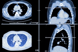
Initial results from a CT lung cancer screening study sponsored by the U.K. National Health Service (NHS) show that low-dose CT exams could detect 70% of lung cancers at stage I or II, according to a report in the Guardian.
The SUMMIT study's principal investigator, Dr. Sam Janes of the University College London Hospitals NHS Foundation Trust (UCLH), told the Guardian that the results represent "a major breakthrough for lung cancer."
"These initial findings, which will be peer-reviewed for publication in a research journal later in the year, highlight the benefits of lung cancer screening for detecting and treating the disease at early stage," UCLH wrote in a post on its website.
Based on these results, CT lung cancer screening could lead to 25% fewer men and 30%-50% fewer women dying from lung cancer, said Dr. Robert Rintoul of the UK Lung Cancer Coalition in the Guardian article. The coalition very much hopes now that a CT lung cancer screening program will be introduced in England, according to Rintoul, who is chair of the coalition's clinical advisory group.
Launched in early 2019, the SUMMIT study aims to detect lung cancer earlier among at-risk Londoners, support the development of a new blood test for early detection of multiple cancers and provide evidence to inform a potential national lung cancer screening program, according to the University College London (UCL) and the University College London Hospitals NHS Foundation Trust.


![Overview of the study design. (A) The fully automated deep learning framework was developed to estimate body composition (BC) (defined as subcutaneous adipose tissue [SAT] in liters; visceral adipose tissue [VAT] in liters; skeletal muscle [SM] in liters; SM fat fraction [SMFF] as a percentage; and intramuscular adipose tissue [IMAT] in deciliters) from MRI. The fully automated framework comprised one model (model 1) to quantify different BC measures (SAT, VAT, SM, SMFF, and IMAT) as three-dimensional (3D) measures from whole-body MRI scans. The second model (model 2) was trained to identify standardized anatomic landmarks along the craniocaudal body axis (z coordinate field), which allowed for subdividing the whole-body measures into different subregions typically examined on clinical routine MRI scans (chest, abdomen, and pelvis). (B) BC was quantified from whole-body MRI in over 66,000 individuals from two large population-based cohort studies, the UK Biobank (UKB) (36,317 individuals) and the German National Cohort (NAKO) (30,291 individuals). Bar graphs show age distribution by sex and cohort. BMI = body mass index. (C) After the performance assessment of the fully automated framework, the change in BC measures, distributions, and profiles across age decades were investigated. Age-, sex-, and height-adjusted body composition reference curves were calculated and made publicly available in a web-based z-score calculator (https://circ-ml.github.io).](https://img.auntminnieeurope.com/mindful/smg/workspaces/default/uploads/2026/05/body-comp.XgAjTfPj1W.jpg?auto=format%2Ccompress&fit=crop&h=112&q=70&w=112)


