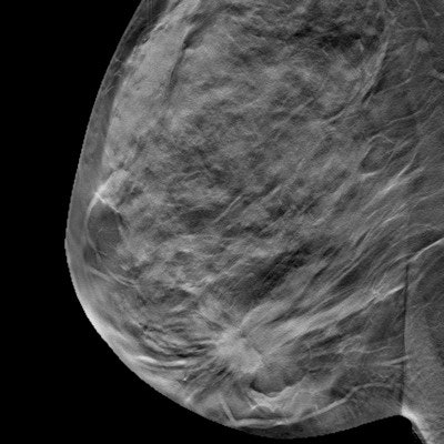
The results of a large, prospective study published on 11 November in Radiology show that digital breast tomosynthesis (DBT) detected more early-stage invasive cancers than full-field digital mammography (FFDM). The study also demonstrated that DBT's benefits extend beyond the initial screening.
DBT has proved its worth as an initial screening tool, but some lingering concerns remain about its efficacy in later screening rounds. The new study puts some of those fears to rest and adds a wealth of prospective research to a topic that has been largely informed by retrospective studies.
"At repeat screening, digital breast tomosynthesis depicts more cancers than digital mammography and finds a lower number of stage II and higher cancers," wrote the team of Italian authors, led by Dr. Francesca Caumo, the director of breast radiology at Veneto Institute of Oncology in Padua. "Such decrease suggests that DBT depicts cancer that might soon become advanced."
The study focused on screening-age women in Verona, Italy, who underwent two rounds of breast screening two years apart. For the first screening round, all 32,870 women in the trial underwent screening with DBT with 2D synthesized mammography (DBT + SM).
However, in a second screening, half the women once again underwent DBT + SM, while the other half received FFDM screening.
The authors compared the trial group's initial and secondary screening data to a third group of 28,680 women screened only with FFDM. Although this control group was not part of the trial, the women still underwent both a baseline and secondary FFDM screening, giving the authors the comparison data they needed for the analysis.
| FFDM vs. DBT + SM by breast screening round | ||||
| Baseline screening | Subsequent screening | |||
| FFDM* | DBT + SM | FFDM* | DBT + SM | |
| Cancer detection rate | 1.06% | 1.97% | 0.5% | 0.8% |
| Recall rate | 11.8% | 6.5% | 3.7% | 3.4% |
| Positive recall rate | 9% | 30.3% | 12% | 23.8% |
Like in previous studies, DBT performed well on the initial screening round. The combination of DBT + SM showed a significant increase in the cancer detection rate and lower recall rate compared with the control group. The group screening with DBT + SM also had a higher positive recall rate.
The benefits of DBT also extended to the subsequent screening round. The group that received DBT + SM for both their first and second screenings had a higher cancer detection rate and a higher positive recall rate than the control group.
But the same wasn't true for the trial group that underwent FFDM for their second screening. This group had a comparable cancer detection rate to the control group as well as a significantly lower positive recall rate.
Perhaps the most notable findings from the study were the effects of DBT on the stage of cancer at detection. In the first round, the women screened with DBT + SM had a higher proportion of stage I cancers and a comparable number of cancers that were stage II or higher.
But in the second round, women screened with DBT + SM in the first round -- regardless of their second-round screening modality -- still had a higher rate of stage I cancer detection than the control group. They also had a lower proportion of stage II cancers than the women only screened with FFDM.
Overall, the findings highlight the potential of DBT to better detect clinically relevant breast cancer beyond the first screening round, according to Dr. Min Sun Bae, an associate professor of radiology at Inha University Hospital and School of Medicine in Incheon, South Korea.
"The results of this study are important because the authors showed that the improved outcomes with DBT are sustainable beyond baseline screening, and that early-stage invasive cancers are more often detected with DBT plus SM than with FFDM at both baseline and subsequent screening," wrote Bae in an associated commentary also published on November 10 in Radiology.
Although the study had a number of merits, the authors were not able to study interval cancers in the study cohort or whether tumor characteristics differed by modality for baseline and subsequent screening. While Bae believes future studies, particularly randomized controlled trials, are needed to corroborate the results, she also said the findings support the growing use of DBT.
"This study supports the routine use of DBT in breast cancer screening for early detection of invasive cancer and improved screening accuracy," she concluded.