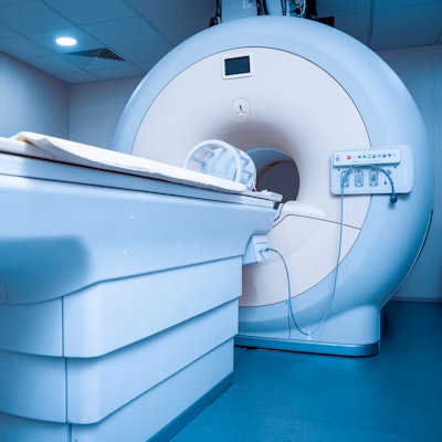
If you could design the MRI scanner of the future, how would you do it? Tomorrow's MRI systems will use advanced coil arrays, novel sensors, new data acquisition techniques, and machine learning, according to a plenary session at last week's International Society for Magnetic Resonance in Medicine (ISMRM) virtual meeting.
The amount of data acquired by MR is increasing, which is a result of a growing number of radiofrequency channels and a trend toward higher acquisition duty cycles in many of today's MR systems.
In the future, MR equipment will be more platform-based, incorporating any number of scanners, explained David Brunner, PhD, chief technology officer and co-founder of Skope Magnetic Resonance Technologies, a Switzerland-based developer of sensor technology and algorithms for the next generation of MRI systems.
The need to improve techniques for acquiring clean MR images is apparent, according to Brunner. He described the optimum MRI platform of the future as being designed with an entirely different infrastructure from what is known today -- one that involves the use of reconfigurable hardware, new interfaces, and real-time processing resources, all aimed at more rapidly processing imaging and machine system operating data.
One of the main deficiencies of MRI to date has been its slow acquisition speed. But Brunner believes that this could be improved by using statistically enhanced stack-on data, based on software modeling and machine learning. Stacking can effectively enhance data signals, and machine-learning software would be trained on the MRI data and then developed into MRI-applicable algorithms and software for later use.
The aim of this approach is to enable MRI systems to learn new information about the biology of patients picked up by the MR scans, according to Brunner. But the information and data required probably cannot be acquired on many of the individual scanners of the kind in use today, he indicated.
To solve this data bottleneck, standalone scanners would need to communicate with other scanners. MRI systems would feature high connectivity and be established on one platform to communicate effectively. The scanners would be fed the same data, and all would interpret the image data in the same manner, giving the data the same meaning consistently.
Brunner has suggested using reconfigurable hardware with a custom-tailored assembly of interfaces and real-time processing resources. Augmenting MRI systems with additional sensors would improve the nature of MRI data and the way it is acquired.
Additional sensors could include motion sensors and magnetic field sensors that report on the state of the patient being scanned and which monitor the operation of the scanner as well, Brunner said. The sensor data could be added to the image output data to help in making a diagnosis.
When designing the MRI platform of the future, developers would have to provide additional space for integrating additional sensors. But it would be difficult to implement all of the dedicated sensor interfaces in an analog state. So, sensors would have to be digitized along with the scanners on the platform. Brunner suggested one option would involve using a 16-channel digitizer to do so.
To incorporate the circuitry further, Brunner offered an integrated solution based on a thumb-sized system-on-chip, which would offer sufficient throughput and reconfigurable hardware-based parallel processing power for difficult applications, such as the high-speed exchange of data.
Future MRI platforms must support the enrichment of MR data, as well as data from other modalities, if needed by the clinician to make a diagnosis. This can be done through the use of additional sensors, machine learning, and a more extensively integrated workflow, Brunner concluded.


![Examples of ultrasound findings and techniques. (A) Images in a 39-year-old male patient with a mass in the left thigh. The mass is heterogeneous on the B-mode US image (compared with the patient in D) and showed increased microvascularity (superb microvascular imaging [SMI]) and shear-wave elastography (SWE) values. Undifferentiated pleomorphic sarcoma was diagnosed at biopsy (with pleomorphic rhabdomyosarcoma in surgical specimen). (B) Images in an 18-year-old male patient with a mass in the left leg. The mass is hypoechoic on the B-mode image, with no other findings suggestive of malignancy. The lesion is in contact with the cortex of the tibia, which is slightly irregular. CT revealed a doubtful anteromedial tibial erosion. The microvascular study demonstrated high vascularization, suggestive of malignancy. Periosteal Ewing sarcoma was diagnosed with both histologic and immunohistochemical confirmation. (C) Images in a 69-year-old female patient with a lump growing on the outside of the left leg. Multiple SWE examinations were performed (please note the high values obtained in the measurements, whereas the color map highlights the stiffness relative to adjacent tissues). SMI showed areas of increased vascularization to target for sampling. Undifferentiated spindle cell sarcoma was diagnosed at biopsy, with residual leiomyosarcoma in the surgical specimen after neoadjuvant therapy. (D) Images in a 56-year-old female patient with a mass in the right thigh. The mass is heterogeneous at both B-mode ultrasound (similar to patient A) and MRI (coronal T2-weighted spectral attenuated inversion recovery [SPAIR]; T1-weighted pre-contrast and postcontrast imaging), which even shows uptake after the administration of paramagnetic contrast material, which is traditionally suggestive of malignancy. Low values at SMI and elastography are suggestive of benignity. Spindle cell lipoma was diagnosed at biopsy, with atypical spindle cell lipomatous tumor in the surgical specimen.](https://img.auntminnieeurope.com/mindful/smg/workspaces/default/uploads/2026/08/images-radiol250278fig2.APCFLSvX6p.jpg?auto=format%2Ccompress&fit=crop&h=100&q=70&w=100)


![Examples of ultrasound findings and techniques. (A) Images in a 39-year-old male patient with a mass in the left thigh. The mass is heterogeneous on the B-mode US image (compared with the patient in D) and showed increased microvascularity (superb microvascular imaging [SMI]) and shear-wave elastography (SWE) values. Undifferentiated pleomorphic sarcoma was diagnosed at biopsy (with pleomorphic rhabdomyosarcoma in surgical specimen). (B) Images in an 18-year-old male patient with a mass in the left leg. The mass is hypoechoic on the B-mode image, with no other findings suggestive of malignancy. The lesion is in contact with the cortex of the tibia, which is slightly irregular. CT revealed a doubtful anteromedial tibial erosion. The microvascular study demonstrated high vascularization, suggestive of malignancy. Periosteal Ewing sarcoma was diagnosed with both histologic and immunohistochemical confirmation. (C) Images in a 69-year-old female patient with a lump growing on the outside of the left leg. Multiple SWE examinations were performed (please note the high values obtained in the measurements, whereas the color map highlights the stiffness relative to adjacent tissues). SMI showed areas of increased vascularization to target for sampling. Undifferentiated spindle cell sarcoma was diagnosed at biopsy, with residual leiomyosarcoma in the surgical specimen after neoadjuvant therapy. (D) Images in a 56-year-old female patient with a mass in the right thigh. The mass is heterogeneous at both B-mode ultrasound (similar to patient A) and MRI (coronal T2-weighted spectral attenuated inversion recovery [SPAIR]; T1-weighted pre-contrast and postcontrast imaging), which even shows uptake after the administration of paramagnetic contrast material, which is traditionally suggestive of malignancy. Low values at SMI and elastography are suggestive of benignity. Spindle cell lipoma was diagnosed at biopsy, with atypical spindle cell lipomatous tumor in the surgical specimen.](https://img.auntminnieeurope.com/mindful/smg/workspaces/default/uploads/2026/08/images-radiol250278fig2.APCFLSvX6p.jpg?auto=format%2Ccompress&fit=crop&h=112&q=70&w=112)