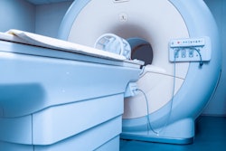
Running an MRI service is like being the captain of a ship on the ocean, according to a keynote presentation at the International Society for Magnetic Resonance in Medicine (ISMRM) virtual conference. Smooth navigation depends on effective collaboration with physicists, technologists, and other staff.
Dr. Pia Sundgren, PhD.Among the required traits for a dedicated MRI radiologist is strict adherence to MR imaging and safety protocols, while balancing department and economics resources for the benefit of both the facility and the patient, said Dr. Pia Sundgren, PhD, professor in the department of clinical sciences and radiology and co-director of the bioimaging center at Lund University in Sweden.
"The radiologist or the [medical doctor] is the captain on the ship when it comes to all the ethical considerations that we have to think about," she said. "We are medically responsible for the patient and we also are responsible to secure the protocols for scanning the patients and patient MR safety."
Among the critical questions is whether a request for an MRI scan is appropriate. To answer this query, radiologists need accurate clinical information to ensure MRI is the best imaging strategy for a particular indication. For one, collaboration between the radiologist, technologist, and referring physician can rule out any contraindications.
"It is important to have clear guidelines for the procedure, from receiving a request, to the protocol in an MRI examination, to performing the study and reading the report," Sundgren told ISMRM's virtual viewers. "We also must have established an MRI safety routine and have a specific MRI radiologist who can take the calls from the MR radiographer, review the MR safety concerns, and handle the MR department resources."
An evaluation of those resources includes comparing the number of available MRI scanners with the number of requested patient scans. A decision can be formulated by the radiologist and physician based on prioritizing the patient's condition to determine which scans are urgent and which scans can wait, she added. Another critical component is reviewing potential MR safety issues with an MR physicist, technologist, or physician to make sure there are no devices in the patient that are not suitable for MR imaging.
"As a radiologist, it is our responsibility to have dedicated MR protocols that are related to the organ of interest, the clinical question, known routines on specific protocols within the department, and protocols that are related to the suspected disease or the suspected diagnosis," Sundgren said. "These protocols must be prescribed and followed to avoid any misunderstanding between the radiologist and the MR radiographer who is performing the examination."
While MRI safety is always a priority, Sundgren said it can be difficult to get radiologists to return for follow-up training sessions or to renew their safety certification once they have completed their initial education, since they spend so little time, if any, in the MRI suite.
"Still, [radiologists] are the ones who have the medical and overall responsibility in an emergency situation, as well as the medical and legal responsibility for what happens to a patient in the MR scanner," she added. "A solution for this could be to require recertification and that there should be e-learning modules available to radiologists, as well as hands-on and personal training with certification on an annual basis, just as they would be for the radiographers and physicists."
Sundgren postulated that radiologists, physicists, and technologists could obtain a "driving license" certification to ensure they can safely operate and oversee the scanner. The certificate would be renewed annually to ensure everyone is up-to-date on latest safety protocols.

![Examples of ultrasound findings and techniques. (A) Images in a 39-year-old male patient with a mass in the left thigh. The mass is heterogeneous on the B-mode US image (compared with the patient in D) and showed increased microvascularity (superb microvascular imaging [SMI]) and shear-wave elastography (SWE) values. Undifferentiated pleomorphic sarcoma was diagnosed at biopsy (with pleomorphic rhabdomyosarcoma in surgical specimen). (B) Images in an 18-year-old male patient with a mass in the left leg. The mass is hypoechoic on the B-mode image, with no other findings suggestive of malignancy. The lesion is in contact with the cortex of the tibia, which is slightly irregular. CT revealed a doubtful anteromedial tibial erosion. The microvascular study demonstrated high vascularization, suggestive of malignancy. Periosteal Ewing sarcoma was diagnosed with both histologic and immunohistochemical confirmation. (C) Images in a 69-year-old female patient with a lump growing on the outside of the left leg. Multiple SWE examinations were performed (please note the high values obtained in the measurements, whereas the color map highlights the stiffness relative to adjacent tissues). SMI showed areas of increased vascularization to target for sampling. Undifferentiated spindle cell sarcoma was diagnosed at biopsy, with residual leiomyosarcoma in the surgical specimen after neoadjuvant therapy. (D) Images in a 56-year-old female patient with a mass in the right thigh. The mass is heterogeneous at both B-mode ultrasound (similar to patient A) and MRI (coronal T2-weighted spectral attenuated inversion recovery [SPAIR]; T1-weighted pre-contrast and postcontrast imaging), which even shows uptake after the administration of paramagnetic contrast material, which is traditionally suggestive of malignancy. Low values at SMI and elastography are suggestive of benignity. Spindle cell lipoma was diagnosed at biopsy, with atypical spindle cell lipomatous tumor in the surgical specimen.](https://img.auntminnieeurope.com/mindful/smg/workspaces/default/uploads/2026/08/images-radiol250278fig2.APCFLSvX6p.jpg?auto=format%2Ccompress&fit=crop&h=100&q=70&w=100)


![Examples of ultrasound findings and techniques. (A) Images in a 39-year-old male patient with a mass in the left thigh. The mass is heterogeneous on the B-mode US image (compared with the patient in D) and showed increased microvascularity (superb microvascular imaging [SMI]) and shear-wave elastography (SWE) values. Undifferentiated pleomorphic sarcoma was diagnosed at biopsy (with pleomorphic rhabdomyosarcoma in surgical specimen). (B) Images in an 18-year-old male patient with a mass in the left leg. The mass is hypoechoic on the B-mode image, with no other findings suggestive of malignancy. The lesion is in contact with the cortex of the tibia, which is slightly irregular. CT revealed a doubtful anteromedial tibial erosion. The microvascular study demonstrated high vascularization, suggestive of malignancy. Periosteal Ewing sarcoma was diagnosed with both histologic and immunohistochemical confirmation. (C) Images in a 69-year-old female patient with a lump growing on the outside of the left leg. Multiple SWE examinations were performed (please note the high values obtained in the measurements, whereas the color map highlights the stiffness relative to adjacent tissues). SMI showed areas of increased vascularization to target for sampling. Undifferentiated spindle cell sarcoma was diagnosed at biopsy, with residual leiomyosarcoma in the surgical specimen after neoadjuvant therapy. (D) Images in a 56-year-old female patient with a mass in the right thigh. The mass is heterogeneous at both B-mode ultrasound (similar to patient A) and MRI (coronal T2-weighted spectral attenuated inversion recovery [SPAIR]; T1-weighted pre-contrast and postcontrast imaging), which even shows uptake after the administration of paramagnetic contrast material, which is traditionally suggestive of malignancy. Low values at SMI and elastography are suggestive of benignity. Spindle cell lipoma was diagnosed at biopsy, with atypical spindle cell lipomatous tumor in the surgical specimen.](https://img.auntminnieeurope.com/mindful/smg/workspaces/default/uploads/2026/08/images-radiol250278fig2.APCFLSvX6p.jpg?auto=format%2Ccompress&fit=crop&h=112&q=70&w=112)