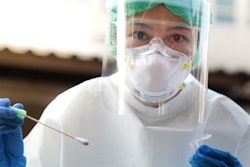
Results from a national survey conducted by the French union of private radiologists (Fédération Nationale de Médecins Radiologues, FNMR) revealed that 77% of private radiologists have received and examined patients who have -- or are suspected of having -- COVID-19. Of the sample, 80% stated that they did not have sufficient personal protective equipment (PPE) for themselves, technicians, and secretaries.
The findings show that the COVID-19 crisis has seriously affected the organization and turnover of all centers, regardless of their size, the FNMR believes. According to the survey, 71% of respondents noted a fall of 80% or more in their activity and the closure of certain sites in order to help others maintain the continuity of patient care and safety.
Despite governmental measures to support businesses and employees, imaging centers must continue to pay charges, according to the FNMR, which pointed to 63% with charges of more than 50,000 euros per month. This coupled with the drop in activity is threatening the existence of dozens of imaging centers, meaning patients may risk losing access to local radiology services.
The union also decried the way some regional health authorities have treated private radiologists, by preventing them from using previously shared hospital-based MRI and CT scanners for their patients. These limitations must cease after the crisis, as much to better fight COVID-19 as to ensure the general population has access to imaging, the FNMR stated. It pointed to its members' readiness to return to normal activity for ambulatory and cancer patients, with organized breast cancer screening a priority.
The FNMR called for compensation to safeguard private imaging, which accounts for up to 80% of imaging in France, and stated that it can no longer accept new economic measures that will weaken the specialty.