
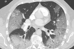
Public health authorities in New South Wales, Australia, believe that a radiology seminar in the country in February could be linked to two cases of the novel coronavirus disease COVID-19 in physicians who tested positive for the virus after the meeting.
Investigators said that 77 physicians attended the seminar on 18 February. A doctor who attended the meeting, a registrar from Liverpool Hospital in the Sydney area, tested positive for coronavirus infection, according to a 4 March news report in the Guardian. The seminar has also been linked to COVID-19 in a doctor at Ryde Hospital, according to the article. Neither of the doctors had traveled overseas or had contact with known COVID-19 patients.
No other attendees at the seminar have shown COVID-19 symptoms, according to authorities. This is a positive sign, as the seminar was more than two weeks ago, and the incubation period for the novel coronavirus that causes COVID-19 is believed to be about two weeks, according to an article on 7news.com.au.
The incident could represent the realization of the worst fears of many public health authorities and care providers: medical workers contracting coronavirus at professional functions.
Indeed, a wide range of medical meetings were canceled this week in an effort to prevent the spread of coronavirus among medical professionals. These include ECR and the annual meetings of the Healthcare Information and Management Systems Society and the Society of Interventional Radiology.


![Overview of the study design. (A) The fully automated deep learning framework was developed to estimate body composition (BC) (defined as subcutaneous adipose tissue [SAT] in liters; visceral adipose tissue [VAT] in liters; skeletal muscle [SM] in liters; SM fat fraction [SMFF] as a percentage; and intramuscular adipose tissue [IMAT] in deciliters) from MRI. The fully automated framework comprised one model (model 1) to quantify different BC measures (SAT, VAT, SM, SMFF, and IMAT) as three-dimensional (3D) measures from whole-body MRI scans. The second model (model 2) was trained to identify standardized anatomic landmarks along the craniocaudal body axis (z coordinate field), which allowed for subdividing the whole-body measures into different subregions typically examined on clinical routine MRI scans (chest, abdomen, and pelvis). (B) BC was quantified from whole-body MRI in over 66,000 individuals from two large population-based cohort studies, the UK Biobank (UKB) (36,317 individuals) and the German National Cohort (NAKO) (30,291 individuals). Bar graphs show age distribution by sex and cohort. BMI = body mass index. (C) After the performance assessment of the fully automated framework, the change in BC measures, distributions, and profiles across age decades were investigated. Age-, sex-, and height-adjusted body composition reference curves were calculated and made publicly available in a web-based z-score calculator (https://circ-ml.github.io).](https://img.auntminnieeurope.com/mindful/smg/workspaces/default/uploads/2026/05/body-comp.XgAjTfPj1W.jpg?auto=format%2Ccompress&fit=crop&h=112&q=70&w=112)


