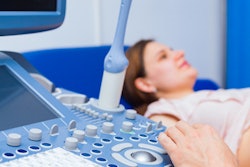
Ultrasound may benefit daily rheumatology practice, according to a study published on 29 January in JCR: Journal of Clinical Rheumatology. Researchers found that ultrasound-guided injections take less than 10 minutes and have a 98% accuracy rate.
Despite studies showing ultrasound's efficacy for some common rheumatological procedures, ultrasound is still not regularly used in many rheumatology departments. The new study suggests ultrasound may be a fast and cost-effective addition to synovial fluid aspirations and drug injections.
"Ultrasound-guided procedures are feasible," wrote the authors, led by Dr. Edoardo Cipolletta from the rheumatology unit at the Carlo Urbani hospital in Jesi, Italy. "In fact, the mean time of the whole procedure was 7 ± 2.5 minutes."
For the study, the authors included 608 rheumatology patients at the Carlo Urbani hospital. Clinicians performed 643 ultrasound-guided procedures, with an average of 5.2 procedures per day. The majority of patients had drug injections or synovial fluid aspiration followed by drug injection.
Ultrasound-guided injections yielded a 98.1% accuracy rate. Injections of the hand, wrist, elbow, and feet had the highest accuracy rate (100%), while shoulder injections had the lowest (92.6%).

The type of ultrasound probe used varied based on the injection site. A medium-frequency probe was used for 73% of procedures, including the majority of knee, shoulder, wrist, ankle, and elbow injections. A high-frequency probe was commonly used for hand and foot procedures, while a convex low-frequency probe was the top choice for hip joint imaging.
Major adverse effects were reported in five patients: two experienced vasovagal reactions, one experienced local skin depigmentation, one had pseudoseptic synovitis, and one had aseptic hip osteonecrosis. No patients experienced an infection that could be related to the use of ultrasound.
"Scientific literature evidence on the risk of infection after musculoskeletal US-guided interventional procedures is scarce," Cipolletta and colleagues wrote.
They noted their study only included one hospital and that not all rheumatology clientele may be able to afford ultrasound equipment. Nevertheless, ultrasound looks like a promising tool for quickly, efficiently, and accurately providing some types of daily rheumatology injections.
"Future research should be focused on the development of an electronic illustrated manual of US-guided musculoskeletal procedures that displays the most relevant information for injecting the principal articular and nonarticular areas of importance in rheumatology, the learning curve of the method, and the development of a shared protocol of disinfection," the authors concluded.


![Examples of ultrasound findings and techniques. (A) Images in a 39-year-old male patient with a mass in the left thigh. The mass is heterogeneous on the B-mode US image (compared with the patient in D) and showed increased microvascularity (superb microvascular imaging [SMI]) and shear-wave elastography (SWE) values. Undifferentiated pleomorphic sarcoma was diagnosed at biopsy (with pleomorphic rhabdomyosarcoma in surgical specimen). (B) Images in an 18-year-old male patient with a mass in the left leg. The mass is hypoechoic on the B-mode image, with no other findings suggestive of malignancy. The lesion is in contact with the cortex of the tibia, which is slightly irregular. CT revealed a doubtful anteromedial tibial erosion. The microvascular study demonstrated high vascularization, suggestive of malignancy. Periosteal Ewing sarcoma was diagnosed with both histologic and immunohistochemical confirmation. (C) Images in a 69-year-old female patient with a lump growing on the outside of the left leg. Multiple SWE examinations were performed (please note the high values obtained in the measurements, whereas the color map highlights the stiffness relative to adjacent tissues). SMI showed areas of increased vascularization to target for sampling. Undifferentiated spindle cell sarcoma was diagnosed at biopsy, with residual leiomyosarcoma in the surgical specimen after neoadjuvant therapy. (D) Images in a 56-year-old female patient with a mass in the right thigh. The mass is heterogeneous at both B-mode ultrasound (similar to patient A) and MRI (coronal T2-weighted spectral attenuated inversion recovery [SPAIR]; T1-weighted pre-contrast and postcontrast imaging), which even shows uptake after the administration of paramagnetic contrast material, which is traditionally suggestive of malignancy. Low values at SMI and elastography are suggestive of benignity. Spindle cell lipoma was diagnosed at biopsy, with atypical spindle cell lipomatous tumor in the surgical specimen.](https://img.auntminnieeurope.com/mindful/smg/workspaces/default/uploads/2026/08/images-radiol250278fig2.APCFLSvX6p.jpg?auto=format%2Ccompress&fit=crop&h=100&q=70&w=100)


![Examples of ultrasound findings and techniques. (A) Images in a 39-year-old male patient with a mass in the left thigh. The mass is heterogeneous on the B-mode US image (compared with the patient in D) and showed increased microvascularity (superb microvascular imaging [SMI]) and shear-wave elastography (SWE) values. Undifferentiated pleomorphic sarcoma was diagnosed at biopsy (with pleomorphic rhabdomyosarcoma in surgical specimen). (B) Images in an 18-year-old male patient with a mass in the left leg. The mass is hypoechoic on the B-mode image, with no other findings suggestive of malignancy. The lesion is in contact with the cortex of the tibia, which is slightly irregular. CT revealed a doubtful anteromedial tibial erosion. The microvascular study demonstrated high vascularization, suggestive of malignancy. Periosteal Ewing sarcoma was diagnosed with both histologic and immunohistochemical confirmation. (C) Images in a 69-year-old female patient with a lump growing on the outside of the left leg. Multiple SWE examinations were performed (please note the high values obtained in the measurements, whereas the color map highlights the stiffness relative to adjacent tissues). SMI showed areas of increased vascularization to target for sampling. Undifferentiated spindle cell sarcoma was diagnosed at biopsy, with residual leiomyosarcoma in the surgical specimen after neoadjuvant therapy. (D) Images in a 56-year-old female patient with a mass in the right thigh. The mass is heterogeneous at both B-mode ultrasound (similar to patient A) and MRI (coronal T2-weighted spectral attenuated inversion recovery [SPAIR]; T1-weighted pre-contrast and postcontrast imaging), which even shows uptake after the administration of paramagnetic contrast material, which is traditionally suggestive of malignancy. Low values at SMI and elastography are suggestive of benignity. Spindle cell lipoma was diagnosed at biopsy, with atypical spindle cell lipomatous tumor in the surgical specimen.](https://img.auntminnieeurope.com/mindful/smg/workspaces/default/uploads/2026/08/images-radiol250278fig2.APCFLSvX6p.jpg?auto=format%2Ccompress&fit=crop&h=112&q=70&w=112)