Investigators from a top Italian facility have employed augmented reality (AR) and 3D printing to plan surgery for a woman with major health complications, including ovarian cancer and an atrial septal defect (ASD). They outlined their experience in a case study published online in JACC: Cardiovascular Interventions.
The researchers from San Donato General Hospital in Milan faced the challenge of restoring adequate blood flow in a 73-year-old woman who presented with shortness of breath and pulmonary hypertension. The patient's medical history included endometrioid ovarian cancer, prior stroke, and class III heart disease. Cardiac ultrasound scans of the patient revealed a sinus venosus ASD that required surgical intervention.
Due to the complexity of the patient's cardiovascular anatomy and pathology, it was critical for the surgical team to understand the precise 3D spatial relationships among all relevant anatomical structures before beginning surgery, first author Dr. Gianfranco Butera, PhD, told AuntMinnieEurope.com.
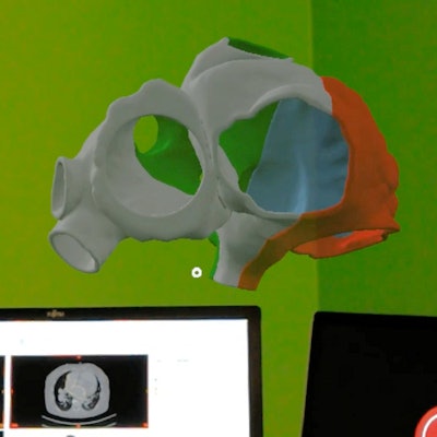
To meet this need, Butera and colleagues generated a virtual 3D model of the patient's heart using computer software. Next, they used an AR headset (HoloLens, Microsoft) to evaluate the hologram and plan their surgical approach (JACC: Cardiovasc Interv, 15 May 2019).
3D cardiac model visualized using augmented reality technology for preoperative planning. Video courtesy of Dr. Gianfranco Butera, PhD.
After studying the individually tailored AR model, the group created a translucent, flexible 3D-printed model of the heart based on conebeam CT scans of the patient. The researchers used the 3D-printed model to test the feasibility of their surgical plan -- in particular, to confirm that the procedure would restore healthy venous blood flow -- after which they performed the operation on the patient. Postoperative ultrasound scans, as well as 3D AR models based on postoperative CT scans, showed significant improvement in patient health.
"Colleagues willing to develop a program about transcatheter treatment of sinus venosus ASD could take advantage of our 3D approach in order to plan successful procedures," Butera said. "The possibility to avoid open heart surgery, cardiac arrest, and extracorporeal circulation is a great advantage for the patients in terms of morbidity, length of hospital stay, [and] pain and discomfort."


![Examples of ultrasound findings and techniques. (A) Images in a 39-year-old male patient with a mass in the left thigh. The mass is heterogeneous on the B-mode US image (compared with the patient in D) and showed increased microvascularity (superb microvascular imaging [SMI]) and shear-wave elastography (SWE) values. Undifferentiated pleomorphic sarcoma was diagnosed at biopsy (with pleomorphic rhabdomyosarcoma in surgical specimen). (B) Images in an 18-year-old male patient with a mass in the left leg. The mass is hypoechoic on the B-mode image, with no other findings suggestive of malignancy. The lesion is in contact with the cortex of the tibia, which is slightly irregular. CT revealed a doubtful anteromedial tibial erosion. The microvascular study demonstrated high vascularization, suggestive of malignancy. Periosteal Ewing sarcoma was diagnosed with both histologic and immunohistochemical confirmation. (C) Images in a 69-year-old female patient with a lump growing on the outside of the left leg. Multiple SWE examinations were performed (please note the high values obtained in the measurements, whereas the color map highlights the stiffness relative to adjacent tissues). SMI showed areas of increased vascularization to target for sampling. Undifferentiated spindle cell sarcoma was diagnosed at biopsy, with residual leiomyosarcoma in the surgical specimen after neoadjuvant therapy. (D) Images in a 56-year-old female patient with a mass in the right thigh. The mass is heterogeneous at both B-mode ultrasound (similar to patient A) and MRI (coronal T2-weighted spectral attenuated inversion recovery [SPAIR]; T1-weighted pre-contrast and postcontrast imaging), which even shows uptake after the administration of paramagnetic contrast material, which is traditionally suggestive of malignancy. Low values at SMI and elastography are suggestive of benignity. Spindle cell lipoma was diagnosed at biopsy, with atypical spindle cell lipomatous tumor in the surgical specimen.](https://img.auntminnieeurope.com/mindful/smg/workspaces/default/uploads/2026/08/images-radiol250278fig2.APCFLSvX6p.jpg?auto=format%2Ccompress&fit=crop&h=100&q=70&w=100)


![Examples of ultrasound findings and techniques. (A) Images in a 39-year-old male patient with a mass in the left thigh. The mass is heterogeneous on the B-mode US image (compared with the patient in D) and showed increased microvascularity (superb microvascular imaging [SMI]) and shear-wave elastography (SWE) values. Undifferentiated pleomorphic sarcoma was diagnosed at biopsy (with pleomorphic rhabdomyosarcoma in surgical specimen). (B) Images in an 18-year-old male patient with a mass in the left leg. The mass is hypoechoic on the B-mode image, with no other findings suggestive of malignancy. The lesion is in contact with the cortex of the tibia, which is slightly irregular. CT revealed a doubtful anteromedial tibial erosion. The microvascular study demonstrated high vascularization, suggestive of malignancy. Periosteal Ewing sarcoma was diagnosed with both histologic and immunohistochemical confirmation. (C) Images in a 69-year-old female patient with a lump growing on the outside of the left leg. Multiple SWE examinations were performed (please note the high values obtained in the measurements, whereas the color map highlights the stiffness relative to adjacent tissues). SMI showed areas of increased vascularization to target for sampling. Undifferentiated spindle cell sarcoma was diagnosed at biopsy, with residual leiomyosarcoma in the surgical specimen after neoadjuvant therapy. (D) Images in a 56-year-old female patient with a mass in the right thigh. The mass is heterogeneous at both B-mode ultrasound (similar to patient A) and MRI (coronal T2-weighted spectral attenuated inversion recovery [SPAIR]; T1-weighted pre-contrast and postcontrast imaging), which even shows uptake after the administration of paramagnetic contrast material, which is traditionally suggestive of malignancy. Low values at SMI and elastography are suggestive of benignity. Spindle cell lipoma was diagnosed at biopsy, with atypical spindle cell lipomatous tumor in the surgical specimen.](https://img.auntminnieeurope.com/mindful/smg/workspaces/default/uploads/2026/08/images-radiol250278fig2.APCFLSvX6p.jpg?auto=format%2Ccompress&fit=crop&h=112&q=70&w=112)