Researchers from Austria are developing a vest embedded with radiofrequency (RF) coils that would allow patients to lie face up during a breast MRI exam -- potentially improving the comfort and accuracy of breast cancer screening.
For standard breast MRI, an individual is required to lie face down inside the MRI scanner, with the breasts fitted into a cup of radiofrequency coils. This process is not only uncomfortable for many people but also makes it difficult for physicians to identify the precise location of tumors on the downward-facing MRI scans during follow-up biopsy, principal investigator Elmar Laistler, PhD, from the Medical University of Vienna said in a statement.
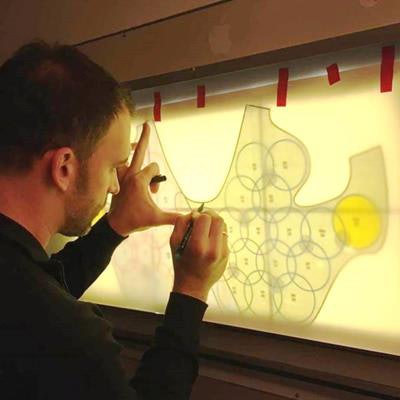
 Pattern for a vest with 32 sewn-in RF coils. Image courtesy of Elmar Laistler, PhD.
In contrast, the group's new technique loads a vest-like device with flexible RF coils that fit on top of the patient, who is then allowed to lie face up inside the MRI scanner during the exam. Early designs of the vest show 32 flexible, 8-cm coils and motion sensors that can correct for image distortions caused by breathing movements.
"An essential aspect of the project is that lying on the back results in the breast being flattened, which means a much larger part of it is close to the receiver," Laistler said. "In this way, the signal is stronger and the measuring time can be shortened."
The Austrian Science Fund (FWF) and French National Research Agency (ANR) are sponsoring the development of the prototype along with its associated motion-correction software.