Talk about a win-win. Choosing either a 1.5-tesla or 3-tesla MRI scan for breast imaging will result in comparably high diagnostic accuracy, according to a study published in the May issue of the European Journal of Radiology.
Austrian and German researchers evaluated images from both magnet strengths and found no statistically significant difference, with 1.5- and 3-tesla breast MR images achieving diagnostic accuracy of approximately 92%. Comparably high numbers were also reached for sensitivity, specificity, and negative predictive values.
"Our findings have several implications: [Breast MRI] is robust and can be performed with various protocols and scanner types," wrote the group led by Dr. Matthias Dietzel from University Hospital Erlangen. "The impact of sequences parameters and the scanner hardware is probably much lower than expected previously. While examinations at 1.5T are cheaper, 3T [breast MRI] has certain inherent advantages: most of all, the potential to reduce the dosage of contrast agents."
The advantages of MRI for breast imaging are well known. The modality provides excellent soft-tissue contrast, multiparametric sequences provide enhanced views of tumor vascularization, and there is simultaneous visualization of both breasts. Multichannel coils can also further aid interpretations of 1.5- and 3-tesla MR images.
Previous studies have compared 1.5- and 3-tesla breast MRI, but much of the research has focused on "small patient groups, specific clinical questions, or selected diagnostic criteria," the authors noted. "Therefore, the evidence, whether increasing the field strength from 1.5T to 3T alters the diagnostic accuracy of breast MRI, is still limited."
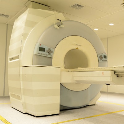
To provide better insight, Dietzel and colleagues reviewed images of 1,961 breasts from 982 patients over a consecutive 12-month period. Of the total, 1,746 breasts (89%) were imaged on a 1.5-tesla scanner (Magnetom Tim Avanto, Siemens Healthineers) and 215 (11%) were imaged on a 3-tesla system (Magnetom Tim Trio, Siemens). Both scanners were equipped with dedicated 16-channel breast coils, and both protocols included contrast-enhanced T1-weighted imaging (EJR, May 2019, Vol. 114, pp. 51-56).
Two readers with expertise in breast MRI assigned a BI-RADS category per breast, with histopathological data or MRI follow-up of more than 24 months serving as the standard of reference.
Overall diagnostic accuracy was 91.9%, with 1.5-tesla MRI at 92% and 3-tesla MRI at 91.2%. Given the spread of only 0.8%, there was no statistically significant difference between the two strengths (p = 0.68). The other diagnostic parameters also did not show a statistically significant difference.
| Diagnostic accuracy based on magnet field strength | ||||
| All cases | 1.5-tesla MRI | 3-tesla MRI | p-value* | |
| Sensitivity | 94.7% | 94.1% | 97.9% | 0.29 |
| Specificity | 91.4% | 91.6% | 89.3% | 0.31 |
| PPV | 66.6% | 65.7% | 71.9% | 0.33 |
| NPV | 99% | 98.9% | 99.3% | 0.65 |
| Accuracy | 91.9% | 92% | 91.2% | 0.68 |
*No statistically significant difference between 1.5 and 3 tesla.
"As a secondary result our data provide evidence that high image quality can be achieved by any MRI unit," the researchers wrote. "To achieve a high image quality, a standard clinical scanner equipped with a breast surface coil is sufficient. The only prerequisite is the optimization of the protocol for the specific scanner hardware (magnet, coil, etc.). This secondary result is important, as there has been much discussion in the past decades which protocol is necessary for accurate diagnosis."


![Examples of ultrasound findings and techniques. (A) Images in a 39-year-old male patient with a mass in the left thigh. The mass is heterogeneous on the B-mode US image (compared with the patient in D) and showed increased microvascularity (superb microvascular imaging [SMI]) and shear-wave elastography (SWE) values. Undifferentiated pleomorphic sarcoma was diagnosed at biopsy (with pleomorphic rhabdomyosarcoma in surgical specimen). (B) Images in an 18-year-old male patient with a mass in the left leg. The mass is hypoechoic on the B-mode image, with no other findings suggestive of malignancy. The lesion is in contact with the cortex of the tibia, which is slightly irregular. CT revealed a doubtful anteromedial tibial erosion. The microvascular study demonstrated high vascularization, suggestive of malignancy. Periosteal Ewing sarcoma was diagnosed with both histologic and immunohistochemical confirmation. (C) Images in a 69-year-old female patient with a lump growing on the outside of the left leg. Multiple SWE examinations were performed (please note the high values obtained in the measurements, whereas the color map highlights the stiffness relative to adjacent tissues). SMI showed areas of increased vascularization to target for sampling. Undifferentiated spindle cell sarcoma was diagnosed at biopsy, with residual leiomyosarcoma in the surgical specimen after neoadjuvant therapy. (D) Images in a 56-year-old female patient with a mass in the right thigh. The mass is heterogeneous at both B-mode ultrasound (similar to patient A) and MRI (coronal T2-weighted spectral attenuated inversion recovery [SPAIR]; T1-weighted pre-contrast and postcontrast imaging), which even shows uptake after the administration of paramagnetic contrast material, which is traditionally suggestive of malignancy. Low values at SMI and elastography are suggestive of benignity. Spindle cell lipoma was diagnosed at biopsy, with atypical spindle cell lipomatous tumor in the surgical specimen.](https://img.auntminnieeurope.com/mindful/smg/workspaces/default/uploads/2026/08/images-radiol250278fig2.APCFLSvX6p.jpg?auto=format%2Ccompress&fit=crop&h=100&q=70&w=100)


![Examples of ultrasound findings and techniques. (A) Images in a 39-year-old male patient with a mass in the left thigh. The mass is heterogeneous on the B-mode US image (compared with the patient in D) and showed increased microvascularity (superb microvascular imaging [SMI]) and shear-wave elastography (SWE) values. Undifferentiated pleomorphic sarcoma was diagnosed at biopsy (with pleomorphic rhabdomyosarcoma in surgical specimen). (B) Images in an 18-year-old male patient with a mass in the left leg. The mass is hypoechoic on the B-mode image, with no other findings suggestive of malignancy. The lesion is in contact with the cortex of the tibia, which is slightly irregular. CT revealed a doubtful anteromedial tibial erosion. The microvascular study demonstrated high vascularization, suggestive of malignancy. Periosteal Ewing sarcoma was diagnosed with both histologic and immunohistochemical confirmation. (C) Images in a 69-year-old female patient with a lump growing on the outside of the left leg. Multiple SWE examinations were performed (please note the high values obtained in the measurements, whereas the color map highlights the stiffness relative to adjacent tissues). SMI showed areas of increased vascularization to target for sampling. Undifferentiated spindle cell sarcoma was diagnosed at biopsy, with residual leiomyosarcoma in the surgical specimen after neoadjuvant therapy. (D) Images in a 56-year-old female patient with a mass in the right thigh. The mass is heterogeneous at both B-mode ultrasound (similar to patient A) and MRI (coronal T2-weighted spectral attenuated inversion recovery [SPAIR]; T1-weighted pre-contrast and postcontrast imaging), which even shows uptake after the administration of paramagnetic contrast material, which is traditionally suggestive of malignancy. Low values at SMI and elastography are suggestive of benignity. Spindle cell lipoma was diagnosed at biopsy, with atypical spindle cell lipomatous tumor in the surgical specimen.](https://img.auntminnieeurope.com/mindful/smg/workspaces/default/uploads/2026/08/images-radiol250278fig2.APCFLSvX6p.jpg?auto=format%2Ccompress&fit=crop&h=112&q=70&w=112)