Dear AuntMinnieEurope Member,
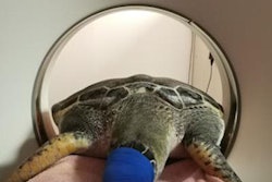
Imaging some humans can be challenging enough, but the logistics of scanning sea creatures are eye-wateringly difficult. The huge variety of species, the virtual lack of protocols and literature, and the need to keep the equipment dry and use sedation are among the numerous challenges.
Valencia in Spain is a center of excellence for imaging of sea life, particularly dolphins, sharks, turtles, walruses, and seahorses. Last Thursday evening, Jose Luis Crespo gave a sparkling talk on this topic, so don't miss this fascinating news report.
Things are getting hot in the EuroMinnies, our new award scheme. The expert panelists have voted, and the list of finalists is now available. We'll name the winners in February, and then present the awards at ECR 2019.
Adrenal imaging is a notoriously tricky area. Incidental adrenal nodules are found in around 5% of patients who undergo CT, and it's vital to keep aware of the full range of pseudolesions and mimics, say radiologists from a top London teaching hospital. They shared their experiences and gave a host of practical tips at RSNA 2018, winning a cum laude award for their efforts.
For patients with chronic low back pain, clinicians often recommend steroid therapy through an image-guided intervention. Swiss researchers have compared the ease of use and reliability of CT- and fluoroscopy-guided spinal injections, and their findings are worth a close look.
Lego MRI came to prominence in 2015 as a way of reassuring children before their scans. A campaign is now underway to convince the Danish company to launch a commercial product, but no less than 10,000 supporters are needed. Read our news report in the MRI Community.
ECR 2019 is less than six weeks away. A major content focus will be on artificial intelligence and interventional radiology, and it also promises to be a significant congress for Italy, which has the presidency. Learn about three new things to see in Vienna.