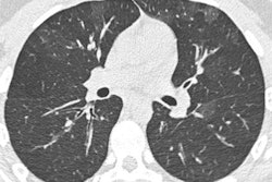
CT lung cancer screening got a big boost this week from researchers in the Dutch-Belgian Randomized Lung Cancer Screening (NELSON) trial, who at the 2018 World Conference on Lung Cancer in Toronto reported a 26% mortality reduction for men in the trial. The findings could give the green light to CT lung screening in Europe.
At the end of the 10-year NELSON study, a randomized controlled trial of CT lung cancer screening in nearly 16,000 eligible participants, CT screening led to a mortality reduction of 26% in men, compared with no screening. The reduction surpasses the 20% found in the National Lung Screening Trial (NLST).
"These findings show that CT screenings are an effective way to assess lung nodules in people at high risk for lung cancer, often leading to detection of suspicious nodules and subsequent surgical intervention at relatively low rates and with few false positives, and can positively increase the chances of cure in this devastating disease," presenter Dr. Harry de Koning, PhD, of Erasmus University Medical Center in Rotterdam told meeting attendees.
Higher mortality reduction
The NELSON CT screening trial is one of the largest of its kind -- second in size only to the NLST. The NLST in 2010 demonstrated that CT lung cancer screening is associated with a statistically significant mortality reduction of 20% from lung cancer and 7% from all causes. Despite the positive findings, most countries beyond the U.S. have yet to implement the test on a national level.
Some skeptics have pointed to potential harms such as overdiagnosis and radiation exposure as reasons not to promote CT screening, and participation rates in the U.S. have, indeed, remained low. Other clinicians have been awaiting further confirmation of the benefits of CT screening from large-scale studies apart from the NLST, notably, the NELSON trial in Europe.
The NELSON trial maintained slightly different criteria for screening and cancer detection from the NLST: The European trial accepted individuals with a slightly younger age and shorter smoking history than the NLST, and it also measured the volume doubling time of lung nodules to determine if the nodules denoted cancer. It also involved three screening exams performed at one, three, and 5.5 years after baseline, in contrast with two annual scans performed each year after a baseline scan.
At the Toronto meeting, de Koning reported on 10-year outcomes of the NELSON trial. The lung cancer mortality reduction from CT screening depended largely on how many rounds of screening individuals went through -- completing all four rounds produced the highest reduction in deaths, he reported.
In total, 250 men died of lung cancer in the control group, as opposed to 157 in the screening arm, indicating a 26% lung cancer mortality reduction from CT screening (p = 0.003). The study was not designed to provide figures on all-cause mortality, de Koning said.
For female participants, lung cancer mortality reduction was even higher: 61% at eight years after baseline screening (p = 0.0037), 53% at nine years (p = 0.0069), and 39% at 10 years (p = 0.0543). The reductions were statistically significant at eight and nine years but not at 10 years.
The NLST was criticized for the large number of suspicious lesions that had to be worked up -- with many turning out to be benign. The NELSON study, however, used a specific follow-up protocol to reduce invasive workup. The 9.3% of cases that were classified as indeterminate received follow-up CT scans at the two-month mark to track nodule volume doubling times, only sending to biopsy those that met established criteria. As a result, only 2.2% of cases turned out to be sent on for further workup, with 0.9% of all cases turning out to be positive for cancer.
These numbers indicate that an individual flagged as having a lung nodule during screening had a 41% risk of actually having lung cancer, de Koning said.
Prime opportunity
What's more, 69% of the screen-detected lung cancers were either stage IA or IB cancers, and only 10% to 12% were stage IV, whereas roughly 50% of the cancers in the control group were stage IV when detected. This difference emphasizes the huge importance of early-stage disease detection offered by CT screening, de Koning noted.
"These results are more favorable than the NLST results, which was already clear-cut, effective, efficient," he said.
When asked what led to the improvement in mortality reduction in the NELSON trial compared with the NLST, de Koning cited several factors that may have contributed to the difference:
- The NLST used x-ray screening for its control group, which itself may have had a mortality benefit.
- The NLST did not incorporate volume doubling time for nodule detection, as NELSON did.
- It had only three rounds of screening, compared with four for NELSON.
The positive NELSON numbers should be a green light to European countries to implement CT lung screening, de Koning said.
"We have shown that even if you are very stringent about referring these men and women, we still have a [mortality] reduction," he said. "So that should be an opportunity for many countries' governments to say that there are two large-scale trials that show large benefits."
Nonetheless, clinicians should be very careful when starting new screening programs, and they should continuously take measures to improve the quality of CT screening in accordance with new research and advances to CT technology, said panelist Dr. Frances Shepherd of Princess Margaret Cancer Centre in Toronto following the presentation.
"There is a steep learning curve for our diagnostic radiologists not to overinterpret," she said. "So these [screening] programs should be implemented quite carefully with strict training and with very strict guidelines for nodule management. ... These screening programs should also go hand in hand with smoking cessation programs."