Dear CT Insider,
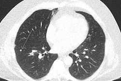
CT lung cancer screening has had a pretty lukewarm response so far across Europe, particularly compared with North America, but evidence about CT's value in this area is now starting to grow.
New cost-effectiveness research from Switzerland deserves close inspection. A team of researchers from Zurich set out to clarify the impact of CT lung screening across a high-income population with a high smoking prevalence, and they found that it can reduce lung cancer mortality by between 6% and 15%. Get the full story here.
In another study published this month, the authors looked at nearly 170,000 children who underwent one or more CT exams in a Dutch hospital between 1979 and 2012. They focused on the prevalence of leukemia and brain tumors, two of the more common malignancies caused by radiation exposure. What exactly did they find? Click here to learn more.
Diagnosis of congenital agenesis of the pericardium can be missed due to the rarity of this cardiac malformation. When cases do arise, CT and MRI can efficiently show the abnormal displacement and rotation of the heart and readily depict absent pericardium, as well as associated cardiac and extracardiac anomalies, Greek researchers said. They shared their experiences at the U.K. Radiological and Radiation Oncology Congress, and you can read more here.
A German group has developed software that uses CT, ultrasound, or MRI scans of the heart to produce virtual 3D models depicting different stages of heartbeat. The researchers unveiled their findings at the Computer Assisted Radiology and Surgery 2018 international congress. Click here for the full story.
Meanwhile, a team led by nuclear medicine expert Dr. Philipp Kaufmann has used software-based cardiac hybrid image fusion to analyze patients who underwent SPECT myocardial perfusion imaging and coronary CT angiography examinations. Click here for our news report.
Finally, a Dutch team examined the brain CT scans of patients with memory complaints and identified a strong association between the presence of calcifications in the hippocampus and various risk factors such as smoking and diabetes. For the details, click here.
This letter highlights just a few of the many articles posted in the CT Community. For the full list, please check out the lineup below.

![Examples of ultrasound findings and techniques. (A) Images in a 39-year-old male patient with a mass in the left thigh. The mass is heterogeneous on the B-mode US image (compared with the patient in D) and showed increased microvascularity (superb microvascular imaging [SMI]) and shear-wave elastography (SWE) values. Undifferentiated pleomorphic sarcoma was diagnosed at biopsy (with pleomorphic rhabdomyosarcoma in surgical specimen). (B) Images in an 18-year-old male patient with a mass in the left leg. The mass is hypoechoic on the B-mode image, with no other findings suggestive of malignancy. The lesion is in contact with the cortex of the tibia, which is slightly irregular. CT revealed a doubtful anteromedial tibial erosion. The microvascular study demonstrated high vascularization, suggestive of malignancy. Periosteal Ewing sarcoma was diagnosed with both histologic and immunohistochemical confirmation. (C) Images in a 69-year-old female patient with a lump growing on the outside of the left leg. Multiple SWE examinations were performed (please note the high values obtained in the measurements, whereas the color map highlights the stiffness relative to adjacent tissues). SMI showed areas of increased vascularization to target for sampling. Undifferentiated spindle cell sarcoma was diagnosed at biopsy, with residual leiomyosarcoma in the surgical specimen after neoadjuvant therapy. (D) Images in a 56-year-old female patient with a mass in the right thigh. The mass is heterogeneous at both B-mode ultrasound (similar to patient A) and MRI (coronal T2-weighted spectral attenuated inversion recovery [SPAIR]; T1-weighted pre-contrast and postcontrast imaging), which even shows uptake after the administration of paramagnetic contrast material, which is traditionally suggestive of malignancy. Low values at SMI and elastography are suggestive of benignity. Spindle cell lipoma was diagnosed at biopsy, with atypical spindle cell lipomatous tumor in the surgical specimen.](https://img.auntminnieeurope.com/mindful/smg/workspaces/default/uploads/2026/08/images-radiol250278fig2.APCFLSvX6p.jpg?auto=format%2Ccompress&fit=crop&h=100&q=70&w=100)





![Examples of ultrasound findings and techniques. (A) Images in a 39-year-old male patient with a mass in the left thigh. The mass is heterogeneous on the B-mode US image (compared with the patient in D) and showed increased microvascularity (superb microvascular imaging [SMI]) and shear-wave elastography (SWE) values. Undifferentiated pleomorphic sarcoma was diagnosed at biopsy (with pleomorphic rhabdomyosarcoma in surgical specimen). (B) Images in an 18-year-old male patient with a mass in the left leg. The mass is hypoechoic on the B-mode image, with no other findings suggestive of malignancy. The lesion is in contact with the cortex of the tibia, which is slightly irregular. CT revealed a doubtful anteromedial tibial erosion. The microvascular study demonstrated high vascularization, suggestive of malignancy. Periosteal Ewing sarcoma was diagnosed with both histologic and immunohistochemical confirmation. (C) Images in a 69-year-old female patient with a lump growing on the outside of the left leg. Multiple SWE examinations were performed (please note the high values obtained in the measurements, whereas the color map highlights the stiffness relative to adjacent tissues). SMI showed areas of increased vascularization to target for sampling. Undifferentiated spindle cell sarcoma was diagnosed at biopsy, with residual leiomyosarcoma in the surgical specimen after neoadjuvant therapy. (D) Images in a 56-year-old female patient with a mass in the right thigh. The mass is heterogeneous at both B-mode ultrasound (similar to patient A) and MRI (coronal T2-weighted spectral attenuated inversion recovery [SPAIR]; T1-weighted pre-contrast and postcontrast imaging), which even shows uptake after the administration of paramagnetic contrast material, which is traditionally suggestive of malignancy. Low values at SMI and elastography are suggestive of benignity. Spindle cell lipoma was diagnosed at biopsy, with atypical spindle cell lipomatous tumor in the surgical specimen.](https://img.auntminnieeurope.com/mindful/smg/workspaces/default/uploads/2026/08/images-radiol250278fig2.APCFLSvX6p.jpg?auto=format%2Ccompress&dpr=2&fit=crop&h=167&q=70&w=250)


