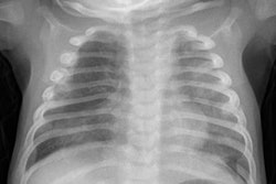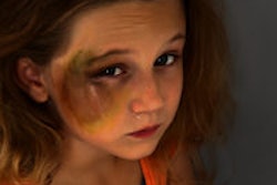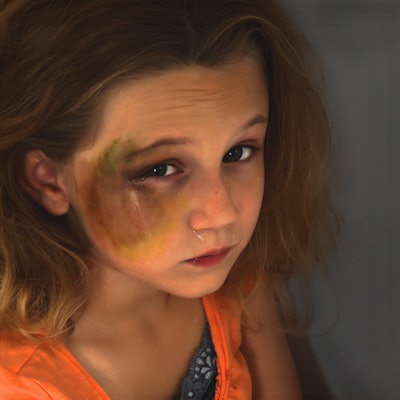
Imaging modalities such as x-ray, CT, and MRI play a key diagnostic role in cases of suspected violence against children. Some findings are made by chance, while others are abundantly apparent. In this interview, Dr. Hans-Joachim Mentzel, professor and head of pediatric radiology at Jena University Hospital's diagnostic and interventional radiology institute in Germany and chair of the German Pediatric Radiology Association, explains the signs he and his colleagues look for.
How often are you confronted with violence against children in your work?
Mentzel: As a pediatric radiologist, you always have it at the back of your mind when you analyze images and happen to notice things. Fortunately, you don't often see children who've been violently shaken and are comatose when they're brought into emergency departments, but when it does happen, you follow a diagnostic imaging checklist. You start with a CT scan so the child gets the right surgery, and then, depending on what you find, you look at the surrounding structures. Choosing the right methods and taking radiological protection into account can be a real challenge.
Dr. Hans-Joachim Mentzel from Jena.We always have to ask ourselves whether broken bones fit with the "accident" that's been described to us, and with the child's age. If we have doubts, we don't take unilateral action; we talk to pediatric physicians and surgeons to get their impression of the child's home environment and the credibility of the information we've been given, and we discuss what to do next. Depending upon the child's age and the relevant guidelines, we'll then carry out a series of high-quality x-rays in the pediatric radiology department to identify or exclude other injuries.
At my hospital, the Thüringen state child protection clinic is right next to the pediatric radiology department, so we don't have far to go and we can discuss things immediately. It's also very important to work with the medicolegal experts who are involved in all physical abuse cases in the state. And we're often contacted by radiologists and child protection departments at other hospitals, so we can agree on common approaches to imaging procedures and evaluation.
In your experience, what kind of injuries are most common in abuse cases?
The injuries we see in radiology depend on the extent of the trauma and the child's age. They range from simple fractures to serious brain damage in shaken infants, including skull fractures, retinal and meningeal bleeds, and brain tissue tears and contusions. These often cause lasting damage to the child.

We carried out a nationwide study a few years ago and found that ribs, arms, and legs are typically broken in specific places, for example as a result of fending off blows or twisting the body away from the attacker. Another typical injury is corner fractures in the growth regions of the bones.
Is there sufficient awareness of the issue in the medical profession and society as a whole, or do you think there needs to be more education?
Even when there's plenty of information available about child abuse in the media and elsewhere, we should never tire of telling people to look out for the signs. We're radiologists, so we can see injuries to the brain, skeleton, and internal organs, but these aren't always immediately obvious from the outside, where there may just be a bruise.
A child who's disturbed or suddenly starts behaving oddly should make us suspicious. All medical professionals need to be constantly aware of the issue. There are plenty of courses for pediatricians, pediatric surgeons, and radiologists, but other groups, such as dentists, should also be taught more about the various aspects of child protection.
Editor's note: This is an edited version of a translation of an article published in German online by the German Radiological Society (DRG, Deutsche Röntgengesellschaft). Translation by Syntacta Translation & Interpreting. To read the original article, click here.



![Examples of ultrasound findings and techniques. (A) Images in a 39-year-old male patient with a mass in the left thigh. The mass is heterogeneous on the B-mode US image (compared with the patient in D) and showed increased microvascularity (superb microvascular imaging [SMI]) and shear-wave elastography (SWE) values. Undifferentiated pleomorphic sarcoma was diagnosed at biopsy (with pleomorphic rhabdomyosarcoma in surgical specimen). (B) Images in an 18-year-old male patient with a mass in the left leg. The mass is hypoechoic on the B-mode image, with no other findings suggestive of malignancy. The lesion is in contact with the cortex of the tibia, which is slightly irregular. CT revealed a doubtful anteromedial tibial erosion. The microvascular study demonstrated high vascularization, suggestive of malignancy. Periosteal Ewing sarcoma was diagnosed with both histologic and immunohistochemical confirmation. (C) Images in a 69-year-old female patient with a lump growing on the outside of the left leg. Multiple SWE examinations were performed (please note the high values obtained in the measurements, whereas the color map highlights the stiffness relative to adjacent tissues). SMI showed areas of increased vascularization to target for sampling. Undifferentiated spindle cell sarcoma was diagnosed at biopsy, with residual leiomyosarcoma in the surgical specimen after neoadjuvant therapy. (D) Images in a 56-year-old female patient with a mass in the right thigh. The mass is heterogeneous at both B-mode ultrasound (similar to patient A) and MRI (coronal T2-weighted spectral attenuated inversion recovery [SPAIR]; T1-weighted pre-contrast and postcontrast imaging), which even shows uptake after the administration of paramagnetic contrast material, which is traditionally suggestive of malignancy. Low values at SMI and elastography are suggestive of benignity. Spindle cell lipoma was diagnosed at biopsy, with atypical spindle cell lipomatous tumor in the surgical specimen.](https://img.auntminnieeurope.com/mindful/smg/workspaces/default/uploads/2026/08/images-radiol250278fig2.APCFLSvX6p.jpg?auto=format%2Ccompress&fit=crop&h=100&q=70&w=100)


![Examples of ultrasound findings and techniques. (A) Images in a 39-year-old male patient with a mass in the left thigh. The mass is heterogeneous on the B-mode US image (compared with the patient in D) and showed increased microvascularity (superb microvascular imaging [SMI]) and shear-wave elastography (SWE) values. Undifferentiated pleomorphic sarcoma was diagnosed at biopsy (with pleomorphic rhabdomyosarcoma in surgical specimen). (B) Images in an 18-year-old male patient with a mass in the left leg. The mass is hypoechoic on the B-mode image, with no other findings suggestive of malignancy. The lesion is in contact with the cortex of the tibia, which is slightly irregular. CT revealed a doubtful anteromedial tibial erosion. The microvascular study demonstrated high vascularization, suggestive of malignancy. Periosteal Ewing sarcoma was diagnosed with both histologic and immunohistochemical confirmation. (C) Images in a 69-year-old female patient with a lump growing on the outside of the left leg. Multiple SWE examinations were performed (please note the high values obtained in the measurements, whereas the color map highlights the stiffness relative to adjacent tissues). SMI showed areas of increased vascularization to target for sampling. Undifferentiated spindle cell sarcoma was diagnosed at biopsy, with residual leiomyosarcoma in the surgical specimen after neoadjuvant therapy. (D) Images in a 56-year-old female patient with a mass in the right thigh. The mass is heterogeneous at both B-mode ultrasound (similar to patient A) and MRI (coronal T2-weighted spectral attenuated inversion recovery [SPAIR]; T1-weighted pre-contrast and postcontrast imaging), which even shows uptake after the administration of paramagnetic contrast material, which is traditionally suggestive of malignancy. Low values at SMI and elastography are suggestive of benignity. Spindle cell lipoma was diagnosed at biopsy, with atypical spindle cell lipomatous tumor in the surgical specimen.](https://img.auntminnieeurope.com/mindful/smg/workspaces/default/uploads/2026/08/images-radiol250278fig2.APCFLSvX6p.jpg?auto=format%2Ccompress&fit=crop&h=112&q=70&w=112)