Researchers in Switzerland are setting their sights on fluoroethyl-tyrosine (FET) PET imaging as the best way to assess adult brainstem glioma and gauge patients' chances for progression-free survival. Study results were presented on Sunday at the Society of Nuclear Medicine and Molecular Imaging (SNMMI) annual meeting.
The group from University Hospital Zurich found that with FET-PET as the guiding modality, patients with a maximum tumor-to-brain ratio (TBRmax) of less than 2.0 had significantly more days of progression-free survival than patients above that 2.0 TBRmax benchmark. FET-PET, however, did not quite pass muster in differentiating between low-grade and high-grade brainstem gliomas.
"Nevertheless, it is interesting to see how FET-PET in this very difficult entity might be a very good diagnostic tool to assess the aggressiveness of adult brainstem gliomas," said attending physician Dr. Irene Burger, who presented the results, filling in for lead author Dr. Abdulrahman Albatly, who serves in the nuclear medicine department of University Hospital Zurich.
Scarce research
Brainstem glioma is a rare disease that affects only 1% to 2% of the adult population, and there is scarce research on the efficacy of FET-PET for this clinical application. At least for now, MRI remains the diagnostic imaging tool of choice for these tumors.
"Although MRI makes advances and new technologies like spectroscopy can reveal more information about tumors, it is still unsatisfactory. It is probably mostly due to the homogeneity and susceptibilities in this area [of the brainstem glioma]," Burger told SNMMI attendees. "But we know that with supratentorial tumors, the grading and treatment plan based on FET is a very promising tool."
In addition, she said, TBRmax has been known to differentiate between low- and high-grade tumors.
This retrospective study was conducted at University Hospital Zurich and included 12 male and four female patients (mean age, 32 years; range, 16-51 years) who underwent FET-PET between 2010 and 2016. Dynamic PET images were acquired 20 minutes after injection of FET (dose, 135 MBq).
All patients had stable disease at the time of their scans. The researchers also recorded maximum standardized uptake values (SUVmax), tumor-to-brain ratios, and the time activity curve of FET-PET. Progression-free survival was determined based on a mean period of 481 days.
For tumor grades, the researchers had biopsy results for nine of the 16 patients that confirmed the presence of gliomas based on histology. MR morphology was available for the other seven patients to differentiate between high- and low-grade gliomas. In all, the researchers were able to confirm nine high-grade gliomas and seven low-grade gliomas.
"Brainstem gliomas are very difficult to biopsy," Burger explained. "Depending on the area where the tumor lies, clinicians are reluctant to get a biopsy. Therefore, seven patients did not get any histological proof."
By the numbers
Kaplan-Meier analysis showed significantly longer progression-free survival for patients whose tumors had a TBRmax of less than 2.0 on FET-PET. These patients had progression-free survival of 665 days (± 32 days), compared with only 220 days (± 39 days) for those with a TBRmax greater than 2.0 (p = 0.000). There was no statistically significant survival difference between high-grade gliomas and low-grade gliomas.
In addition, SUVmax and TBRmax did not differ significantly between high-grade gliomas and low-grade gliomas (p = 0.711 and 0.672, respectively). Progressive gliomas, however, had significantly higher SUVmax (p = 0.003) and TBRmax (p = 0.001), compared with stable gliomas.
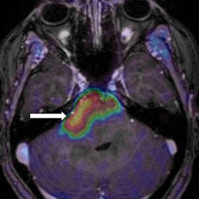
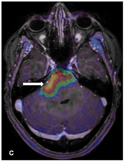
 Contrast enhancement from the PET radiotracer FET highlights the brainstem glioma. Image courtesy of Albatly et al and the Journal of Nuclear Medicine.
Burger cited a few limitations of the study, including the small patient sample. The results have to be "taken with care, the Kaplan-Meier analysis especially in such a small cohort," she said.


