Dear AuntMinnieEurope Member,
The raging controversy surrounding overdiagnosis in screening mammography persisted throughout 2016, and it's showing no signs of subsiding.
Researchers from the Nordic Cochrane Centre in Copenhagen have voiced new concerns over whether screening provides the promised benefits of reduced mortality, less invasive treatment, and reduced disease burden. They say 17 years of organized breast screening in Denmark has not cut the incidence of advanced tumors but has increased the incidence of nonadvanced tumors and ductal carcinoma in situ. Read more in the Women's Imaging Community, or by clicking here.
Also, don't miss our thought-provoking column by a radiologist who was diagnosed with breast cancer at the age of 38. What lessons did Dr. Jennifer Adams learn from her experience as a patient? How has it helped her clinical practice? Click here to find out.
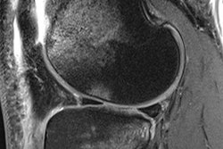
Greater speed in MRI can lead to easier visualization of pathologies and functional anatomy, better imaging within shorter time slots, improved patient care, and increased productivity, according to Swiss radiologists. They think accelerated MRI, which combines dedicated hardware and parallel imaging algorithms, has been developed and improved over the last three decades. Learn about their work in the MRI Community, or click here.
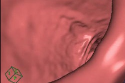
Meanwhile, Italian researchers have joined up with Japanese and U.S. colleagues to participate in a new study on CT colonography. Their findings were presented at RSNA 2016 and are worth a close look. Click here to do so.
Gynecology cases often involve multimodality imaging, which can be complex, and this prompted the gynecology oncology tumor board at London's Imperial College to offer some practical advice at the same Chicago meeting. To discover what the group had to say, click here.


![Examples of ultrasound findings and techniques. (A) Images in a 39-year-old male patient with a mass in the left thigh. The mass is heterogeneous on the B-mode US image (compared with the patient in D) and showed increased microvascularity (superb microvascular imaging [SMI]) and shear-wave elastography (SWE) values. Undifferentiated pleomorphic sarcoma was diagnosed at biopsy (with pleomorphic rhabdomyosarcoma in surgical specimen). (B) Images in an 18-year-old male patient with a mass in the left leg. The mass is hypoechoic on the B-mode image, with no other findings suggestive of malignancy. The lesion is in contact with the cortex of the tibia, which is slightly irregular. CT revealed a doubtful anteromedial tibial erosion. The microvascular study demonstrated high vascularization, suggestive of malignancy. Periosteal Ewing sarcoma was diagnosed with both histologic and immunohistochemical confirmation. (C) Images in a 69-year-old female patient with a lump growing on the outside of the left leg. Multiple SWE examinations were performed (please note the high values obtained in the measurements, whereas the color map highlights the stiffness relative to adjacent tissues). SMI showed areas of increased vascularization to target for sampling. Undifferentiated spindle cell sarcoma was diagnosed at biopsy, with residual leiomyosarcoma in the surgical specimen after neoadjuvant therapy. (D) Images in a 56-year-old female patient with a mass in the right thigh. The mass is heterogeneous at both B-mode ultrasound (similar to patient A) and MRI (coronal T2-weighted spectral attenuated inversion recovery [SPAIR]; T1-weighted pre-contrast and postcontrast imaging), which even shows uptake after the administration of paramagnetic contrast material, which is traditionally suggestive of malignancy. Low values at SMI and elastography are suggestive of benignity. Spindle cell lipoma was diagnosed at biopsy, with atypical spindle cell lipomatous tumor in the surgical specimen.](https://img.auntminnieeurope.com/mindful/smg/workspaces/default/uploads/2026/08/images-radiol250278fig2.APCFLSvX6p.jpg?auto=format%2Ccompress&fit=crop&h=100&q=70&w=100)


![Examples of ultrasound findings and techniques. (A) Images in a 39-year-old male patient with a mass in the left thigh. The mass is heterogeneous on the B-mode US image (compared with the patient in D) and showed increased microvascularity (superb microvascular imaging [SMI]) and shear-wave elastography (SWE) values. Undifferentiated pleomorphic sarcoma was diagnosed at biopsy (with pleomorphic rhabdomyosarcoma in surgical specimen). (B) Images in an 18-year-old male patient with a mass in the left leg. The mass is hypoechoic on the B-mode image, with no other findings suggestive of malignancy. The lesion is in contact with the cortex of the tibia, which is slightly irregular. CT revealed a doubtful anteromedial tibial erosion. The microvascular study demonstrated high vascularization, suggestive of malignancy. Periosteal Ewing sarcoma was diagnosed with both histologic and immunohistochemical confirmation. (C) Images in a 69-year-old female patient with a lump growing on the outside of the left leg. Multiple SWE examinations were performed (please note the high values obtained in the measurements, whereas the color map highlights the stiffness relative to adjacent tissues). SMI showed areas of increased vascularization to target for sampling. Undifferentiated spindle cell sarcoma was diagnosed at biopsy, with residual leiomyosarcoma in the surgical specimen after neoadjuvant therapy. (D) Images in a 56-year-old female patient with a mass in the right thigh. The mass is heterogeneous at both B-mode ultrasound (similar to patient A) and MRI (coronal T2-weighted spectral attenuated inversion recovery [SPAIR]; T1-weighted pre-contrast and postcontrast imaging), which even shows uptake after the administration of paramagnetic contrast material, which is traditionally suggestive of malignancy. Low values at SMI and elastography are suggestive of benignity. Spindle cell lipoma was diagnosed at biopsy, with atypical spindle cell lipomatous tumor in the surgical specimen.](https://img.auntminnieeurope.com/mindful/smg/workspaces/default/uploads/2026/08/images-radiol250278fig2.APCFLSvX6p.jpg?auto=format%2Ccompress&fit=crop&h=112&q=70&w=112)