Dear Cardiac Imaging Insider,
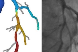
Fractional flow reserve CT (FFR-CT) has already demonstrated strong results in a few trials for its ability to noninvasively confirm obstructive coronary disease. But in one of the most difficult patient groups in the healthcare system -- stable chest-pain patients with an intermediate risk of disease -- a new study found FFR-CT results approaching the level of spectacular.
In a presentation at the recent RSNA 2016 meeting in Chicago, Belgian researchers examined the use of FFR-CT in this group of patients, the care of which requires so much time, energy, and costs in the emergency setting. They found that FFR-CT can rule out a high percentage of false positives by determining almost every time whether a coronary stenosis is hemodynamically significant. Its use could potentially transform the diagnostic paradigm for coronary disease workup if tough political obstacles can be overcome. Learn more by clicking here.
Speaking of chest pain, the U.K. has just released new guidelines for diagnosis and management of the condition, highlighting the role of advanced imaging modalities such as coronary CT angiography, which is now recommended as a first-line test for patients with chest pain. The results pose challenges for even higher-volume CT centers, according to the report you'll find here.
In a second European study presented at RSNA 2016, German radiologists used MRI to determine the effects of short-term sleep deprivation on the heart health of young radiologists, a group that tends to know a thing or two about the topic. The results weren't pretty: The investigators found adverse changes in multiple heart function parameters as sleep levels decreased.
"This is a study that is close to my heart, as I spent a good deal of my life staying awake in residency, but it affects more than just hospital staff," said study author Dr. Suzanne Steinbaum. Get the rest of the story here.
But if sleep loss is bad for the heart, a drink or two is apparently OK, concluded a study from Budapest presented at RSNA 2016. The study used coronary CT angiography to analyze both drinkers and nondrinkers to understand how alcohol may or may not contribute to the development of fatty plaques in the arteries. You'll find the intriguing results by clicking here.
If radiologists live and breathe dose awareness every day, cardiologists as a group have perhaps been slower to embrace the concept of optimizing dose with every exam. But no longer. New efforts are underway to increase dose awareness among European cardiologists, according to a new editorial in the British Journal of Radiology. The article illustrates the interplay between cardiology and radiological techniques, cardiology training, and ionizing radiation regulations, as well as establishes a context for training and accreditation of cardiologists who use radiation.
But not all cardiologists are on board, according to a German cardiologist. At the conference of the British Institute of Radiology last month, he explained why.
We invite you to scroll though the links below for more important news from about cardiac imaging from Europe, conveniently located right here in your Cardiac Imaging Community.


![Examples of ultrasound findings and techniques. (A) Images in a 39-year-old male patient with a mass in the left thigh. The mass is heterogeneous on the B-mode US image (compared with the patient in D) and showed increased microvascularity (superb microvascular imaging [SMI]) and shear-wave elastography (SWE) values. Undifferentiated pleomorphic sarcoma was diagnosed at biopsy (with pleomorphic rhabdomyosarcoma in surgical specimen). (B) Images in an 18-year-old male patient with a mass in the left leg. The mass is hypoechoic on the B-mode image, with no other findings suggestive of malignancy. The lesion is in contact with the cortex of the tibia, which is slightly irregular. CT revealed a doubtful anteromedial tibial erosion. The microvascular study demonstrated high vascularization, suggestive of malignancy. Periosteal Ewing sarcoma was diagnosed with both histologic and immunohistochemical confirmation. (C) Images in a 69-year-old female patient with a lump growing on the outside of the left leg. Multiple SWE examinations were performed (please note the high values obtained in the measurements, whereas the color map highlights the stiffness relative to adjacent tissues). SMI showed areas of increased vascularization to target for sampling. Undifferentiated spindle cell sarcoma was diagnosed at biopsy, with residual leiomyosarcoma in the surgical specimen after neoadjuvant therapy. (D) Images in a 56-year-old female patient with a mass in the right thigh. The mass is heterogeneous at both B-mode ultrasound (similar to patient A) and MRI (coronal T2-weighted spectral attenuated inversion recovery [SPAIR]; T1-weighted pre-contrast and postcontrast imaging), which even shows uptake after the administration of paramagnetic contrast material, which is traditionally suggestive of malignancy. Low values at SMI and elastography are suggestive of benignity. Spindle cell lipoma was diagnosed at biopsy, with atypical spindle cell lipomatous tumor in the surgical specimen.](https://img.auntminnieeurope.com/mindful/smg/workspaces/default/uploads/2026/08/images-radiol250278fig2.APCFLSvX6p.jpg?auto=format%2Ccompress&fit=crop&h=100&q=70&w=100)


![Examples of ultrasound findings and techniques. (A) Images in a 39-year-old male patient with a mass in the left thigh. The mass is heterogeneous on the B-mode US image (compared with the patient in D) and showed increased microvascularity (superb microvascular imaging [SMI]) and shear-wave elastography (SWE) values. Undifferentiated pleomorphic sarcoma was diagnosed at biopsy (with pleomorphic rhabdomyosarcoma in surgical specimen). (B) Images in an 18-year-old male patient with a mass in the left leg. The mass is hypoechoic on the B-mode image, with no other findings suggestive of malignancy. The lesion is in contact with the cortex of the tibia, which is slightly irregular. CT revealed a doubtful anteromedial tibial erosion. The microvascular study demonstrated high vascularization, suggestive of malignancy. Periosteal Ewing sarcoma was diagnosed with both histologic and immunohistochemical confirmation. (C) Images in a 69-year-old female patient with a lump growing on the outside of the left leg. Multiple SWE examinations were performed (please note the high values obtained in the measurements, whereas the color map highlights the stiffness relative to adjacent tissues). SMI showed areas of increased vascularization to target for sampling. Undifferentiated spindle cell sarcoma was diagnosed at biopsy, with residual leiomyosarcoma in the surgical specimen after neoadjuvant therapy. (D) Images in a 56-year-old female patient with a mass in the right thigh. The mass is heterogeneous at both B-mode ultrasound (similar to patient A) and MRI (coronal T2-weighted spectral attenuated inversion recovery [SPAIR]; T1-weighted pre-contrast and postcontrast imaging), which even shows uptake after the administration of paramagnetic contrast material, which is traditionally suggestive of malignancy. Low values at SMI and elastography are suggestive of benignity. Spindle cell lipoma was diagnosed at biopsy, with atypical spindle cell lipomatous tumor in the surgical specimen.](https://img.auntminnieeurope.com/mindful/smg/workspaces/default/uploads/2026/08/images-radiol250278fig2.APCFLSvX6p.jpg?auto=format%2Ccompress&fit=crop&h=112&q=70&w=112)