Dear Cardiac Imaging Insider,
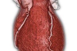
A raft of new studies on coronary CT angiography (CCTA) is refining the use of the powerful modality in patients with suspected coronary disease, and the studies are showing more about how it works in combination with other diagnostic tests to optimize safety, costs, and patient outcomes.
To that end, researchers in Germany compared CCTA directly with cardiac catheterization to look at diagnostic yields, major complications, and adverse events years later. CT was orders of magnitude more efficient in most categories, except radiation dose. Get the rest of the story here.
Meanwhile, Italian investigators wondered if they could zero in on which asymptomatic individuals at risk of heart attack should be screened with CCTA, considering that not everyone can or should undergo a CT scan owing to its cost and radiation dose. Their study looked at C-reactive protein, interleukins, and even the use of carotid ultrasound in an effort to maximize the likelihood that when CT is undertaken it turns out positive. Their innovative study revealed the surprising strength of those other risk factors.
Not to be left out, Austrian researchers analyzed the nearly universal use of degree of stenosis to define coronary artery disease, examining 160 patients with CCTA and invasive coronary angiography. They followed them to measure the predictive value of percent stenosis versus absolute lumen diameter. The results were surprising, as you'll see by clicking here.
How about using cardiac MRI to trim the use of invasive angiography? Researchers in the U.K. looked at more than 1,000 patients and concluded that a cardiac MR-based strategy for assessing coronary disease could significantly reduce need for the invasive procedure. Find the rest of the article here.
AuntMinnieEurope.com contributing writer Dr. Anna Fustier left behind all creature comforts for a six-month assignment in a tiny village in Madagascar. Life may be primitive, and equipment may be scarce, but on the positive side, patients never have a train to catch, and they're never overweight, so ultrasound works beautifully. Learn the rest of the details here.
In other news, French radiologists are aiming to safeguard their profession with the publication of the White Book of radiology earlier this month. Among other things, it promotes pricing that favors the use of noninvasive cardiac imaging exams, as you'll see here.
And a Finnish team investigated how the heart fares when women undergo radiotherapy of the breast. They used echocardiography to pinpoint regional changes in cardiac morphology and function that depended on whether the right or left breast was treated.
For more heart imaging news, we invite you to scroll through the links below, here in your Cardiac Imaging Community.


![Examples of ultrasound findings and techniques. (A) Images in a 39-year-old male patient with a mass in the left thigh. The mass is heterogeneous on the B-mode US image (compared with the patient in D) and showed increased microvascularity (superb microvascular imaging [SMI]) and shear-wave elastography (SWE) values. Undifferentiated pleomorphic sarcoma was diagnosed at biopsy (with pleomorphic rhabdomyosarcoma in surgical specimen). (B) Images in an 18-year-old male patient with a mass in the left leg. The mass is hypoechoic on the B-mode image, with no other findings suggestive of malignancy. The lesion is in contact with the cortex of the tibia, which is slightly irregular. CT revealed a doubtful anteromedial tibial erosion. The microvascular study demonstrated high vascularization, suggestive of malignancy. Periosteal Ewing sarcoma was diagnosed with both histologic and immunohistochemical confirmation. (C) Images in a 69-year-old female patient with a lump growing on the outside of the left leg. Multiple SWE examinations were performed (please note the high values obtained in the measurements, whereas the color map highlights the stiffness relative to adjacent tissues). SMI showed areas of increased vascularization to target for sampling. Undifferentiated spindle cell sarcoma was diagnosed at biopsy, with residual leiomyosarcoma in the surgical specimen after neoadjuvant therapy. (D) Images in a 56-year-old female patient with a mass in the right thigh. The mass is heterogeneous at both B-mode ultrasound (similar to patient A) and MRI (coronal T2-weighted spectral attenuated inversion recovery [SPAIR]; T1-weighted pre-contrast and postcontrast imaging), which even shows uptake after the administration of paramagnetic contrast material, which is traditionally suggestive of malignancy. Low values at SMI and elastography are suggestive of benignity. Spindle cell lipoma was diagnosed at biopsy, with atypical spindle cell lipomatous tumor in the surgical specimen.](https://img.auntminnieeurope.com/mindful/smg/workspaces/default/uploads/2026/08/images-radiol250278fig2.APCFLSvX6p.jpg?auto=format%2Ccompress&fit=crop&h=100&q=70&w=100)


![Examples of ultrasound findings and techniques. (A) Images in a 39-year-old male patient with a mass in the left thigh. The mass is heterogeneous on the B-mode US image (compared with the patient in D) and showed increased microvascularity (superb microvascular imaging [SMI]) and shear-wave elastography (SWE) values. Undifferentiated pleomorphic sarcoma was diagnosed at biopsy (with pleomorphic rhabdomyosarcoma in surgical specimen). (B) Images in an 18-year-old male patient with a mass in the left leg. The mass is hypoechoic on the B-mode image, with no other findings suggestive of malignancy. The lesion is in contact with the cortex of the tibia, which is slightly irregular. CT revealed a doubtful anteromedial tibial erosion. The microvascular study demonstrated high vascularization, suggestive of malignancy. Periosteal Ewing sarcoma was diagnosed with both histologic and immunohistochemical confirmation. (C) Images in a 69-year-old female patient with a lump growing on the outside of the left leg. Multiple SWE examinations were performed (please note the high values obtained in the measurements, whereas the color map highlights the stiffness relative to adjacent tissues). SMI showed areas of increased vascularization to target for sampling. Undifferentiated spindle cell sarcoma was diagnosed at biopsy, with residual leiomyosarcoma in the surgical specimen after neoadjuvant therapy. (D) Images in a 56-year-old female patient with a mass in the right thigh. The mass is heterogeneous at both B-mode ultrasound (similar to patient A) and MRI (coronal T2-weighted spectral attenuated inversion recovery [SPAIR]; T1-weighted pre-contrast and postcontrast imaging), which even shows uptake after the administration of paramagnetic contrast material, which is traditionally suggestive of malignancy. Low values at SMI and elastography are suggestive of benignity. Spindle cell lipoma was diagnosed at biopsy, with atypical spindle cell lipomatous tumor in the surgical specimen.](https://img.auntminnieeurope.com/mindful/smg/workspaces/default/uploads/2026/08/images-radiol250278fig2.APCFLSvX6p.jpg?auto=format%2Ccompress&fit=crop&h=112&q=70&w=112)