PET/MRI is just as proficient as PET/CT in detecting primary cancer and metastases from an unknown primary tumor, making PET/MRI a "powerful alternative" with significantly less exposure of patients to ionizing radiation, according to a German study in the November issue of the European Journal of Radiology.
Researchers from the University Duisburg-Essen and University of Dusseldorf also found that PET/MRI achieved "comparably high" lesion conspicuity and diagnostic confidence results and "superior assessment" of cervical lesions in the comparison with PET/CT.
"Considering the significantly lower dose of ionizing radiation, PET/MRI may serve as a powerful alternative to PET/CT in the future, particularly for therapy monitoring and/or surveillance," wrote lead author Dr. Verena Ruhlmann and colleagues (EJR, November 2016, Vol. 85, pp. 1941-1947).
Diagnostic challenge
Traditionally, clinicians have relied on a myriad of techniques to detect cervical lymph node metastases from an unknown primary tumor. Among the choices are endoscopy of the upper aerodigestive tract, CT and/or MRI of the head and neck, and fine-needle biopsy of the enlarged lymph nodes.
Of course, PET/CT has been a solid mainstay for cancer detection. The authors cited one study that credited the hybrid modality with detecting as many as 58% of primary tumors in patients with suspected metastases from an unknown primary.
With the increasing use of whole-body PET/MRI scanners, Ruhlmann and colleagues chose to explore the combination of functional MRI and metabolic PET data to detect primary cancer and its metastases in patients suspected of cancer from an unknown primary tumor.
The researchers enrolled 15 men and five women (age 53 years, ± 13 years) who were suspected for cancer of unknown primary tumor. The 20 subjects were scheduled for a whole-body FDG-PET/CT scan and then prospectively enrolled for a subsequent PET/MRI exam.
Patients initially underwent a dedicated head and neck PET/CT scan (Biograph mCT, Siemens Healthineers) of 60 minutes duration (± 5 minutes) after a weight-based FDG injection and administration of CT contrast (Ultravist, Bayer HealthCare Pharmaceuticals) 40 seconds later.
After the PET/CT exams, subjects then underwent PET/MRI on an integrated 3-tesla MRI and PET system (Biograph mMR, Siemens). PET mages were acquired in five bed positions from the head to midthigh with the aid of 3D image reconstruction.
Two readers with more than six years' experience in MRI and hybrid imaging separately and randomly interpreted results using a dedicated viewer. Their task included the detection of the primary cancer and metastases, rating lesion conspicuity on a four-point scale, and grading diagnostic confidence on a three-point scale. PET results also were analyzed for maximum standardized uptake values (SUVmax) of all PET-positive lesions using volume of interest from the PET/CT and PET/MR datasets.
Tumor discoveries
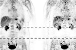
In their final analysis, a total of 49 malignant lesions were discovered in 14 out of 20 patients. PET/CT and PET/MRI both correctly detected the primary cancer in 11 patients (55%), leaving the other nine subjects with no evidence of a primary tumor.
"Our results go inline with previous studies, in revealing an improved identification rate of the primary tumor based on PET/CT and/or PET/MRI of approximately 55% of the cases, which is superior to previously published detection rates derived from conventional imaging," the authors noted.
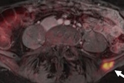
Both hybrid modalities achieved similar results regarding the discovery of metastases. PET/CT detected a total of 38 such abnormalities, compared with 37 identifications made by PET/MRI. The difference was PET/MRI missing one lung metastases in the right upper lobe. Both modalities also correctly concluded six patients (30%) had no metastases.
| Comparison of PET/CT vs. PET/MRI for metastases detection | |||
| PET/CT | PET/MRI | P value | |
| Total abnormalities found | 37 | 38 | |
| Lesion conspicuity | 2.62 ± 0.64 | 2.60 ± 0.64 | |
| Cervical lesions | 1.86 ± 0.90 | 2.43 ± 0.53 | 0.30 |
| Pulmonary lesions | 2.88 ± 0.36 | 2.13 ± 0.99 | 0.10 |
| Diagnostic confidence | 2.74 (± 0.48) | 2.73 (± 0.49) | |
| Mean SUVmax values | 7.2 ± 3.5 | 7.9 ± 4.2 | |
Overall lesion conspicuity between PET/CT (2.62 ± 0.64) and PET/MRI (2.60 ± 0.64) also was comparably high, with a few exceptions. PET/MRI was superior in the assessment of cervical lesions (2.43 ± 0.53 versus 1.86 ± 0.90 for PET/CT). PET/CT, however, was more proficient for pulmonary lesions (2.88 ± 0.36), compared with PET/MRI (2.13 ± 0.99).
"Despite the improved lesion conspicuity of cervical lesions in PET/MRI, the ratings lacked statistical significance, which may be caused by an underpowered analysis due to the small patient cohort," the authors noted.
As for diagnostic confidence, the two hybrid modalities received high marks with PET/CT at 2.74 (± 0.48) and PET/MRI with 2.73 (± 0.49). The mean values of SUVmax of all PET-positive lesions (PET/MRI 7.9 ± 4.2 versus PET/CT 7.2 ± 3.5) showed a strong positive correlation between PET/CT and PET/MR-derived SUVmax.
"Our study demonstrates a comparable detection of primary cancer and metastases in patients suspected for cancer of unknown primary tumor in both PET/CT and PET/MRI," the authors wrote, "with comparably high lesion conspicuity and diagnostic confidence, with indicated (yet not statistically significant) superior assessment of cervical lesions in PET/MRI and pulmonary lesions in PET/CT."
With the diagnostic capabilities of both hybrid imaging techniques comparable in the detection of primary tumor and metastastic spread, one major advantage is significantly reduced radiation exposure from PET/MRI. In this paper, researchers cited several previous studies that estimated a potential ionizing radiation reduction of as much as 75% with PET/MRI compared to PET/CT.

![Examples of ultrasound findings and techniques. (A) Images in a 39-year-old male patient with a mass in the left thigh. The mass is heterogeneous on the B-mode US image (compared with the patient in D) and showed increased microvascularity (superb microvascular imaging [SMI]) and shear-wave elastography (SWE) values. Undifferentiated pleomorphic sarcoma was diagnosed at biopsy (with pleomorphic rhabdomyosarcoma in surgical specimen). (B) Images in an 18-year-old male patient with a mass in the left leg. The mass is hypoechoic on the B-mode image, with no other findings suggestive of malignancy. The lesion is in contact with the cortex of the tibia, which is slightly irregular. CT revealed a doubtful anteromedial tibial erosion. The microvascular study demonstrated high vascularization, suggestive of malignancy. Periosteal Ewing sarcoma was diagnosed with both histologic and immunohistochemical confirmation. (C) Images in a 69-year-old female patient with a lump growing on the outside of the left leg. Multiple SWE examinations were performed (please note the high values obtained in the measurements, whereas the color map highlights the stiffness relative to adjacent tissues). SMI showed areas of increased vascularization to target for sampling. Undifferentiated spindle cell sarcoma was diagnosed at biopsy, with residual leiomyosarcoma in the surgical specimen after neoadjuvant therapy. (D) Images in a 56-year-old female patient with a mass in the right thigh. The mass is heterogeneous at both B-mode ultrasound (similar to patient A) and MRI (coronal T2-weighted spectral attenuated inversion recovery [SPAIR]; T1-weighted pre-contrast and postcontrast imaging), which even shows uptake after the administration of paramagnetic contrast material, which is traditionally suggestive of malignancy. Low values at SMI and elastography are suggestive of benignity. Spindle cell lipoma was diagnosed at biopsy, with atypical spindle cell lipomatous tumor in the surgical specimen.](https://img.auntminnieeurope.com/mindful/smg/workspaces/default/uploads/2026/08/images-radiol250278fig2.APCFLSvX6p.jpg?auto=format%2Ccompress&fit=crop&h=100&q=70&w=100)



![Examples of ultrasound findings and techniques. (A) Images in a 39-year-old male patient with a mass in the left thigh. The mass is heterogeneous on the B-mode US image (compared with the patient in D) and showed increased microvascularity (superb microvascular imaging [SMI]) and shear-wave elastography (SWE) values. Undifferentiated pleomorphic sarcoma was diagnosed at biopsy (with pleomorphic rhabdomyosarcoma in surgical specimen). (B) Images in an 18-year-old male patient with a mass in the left leg. The mass is hypoechoic on the B-mode image, with no other findings suggestive of malignancy. The lesion is in contact with the cortex of the tibia, which is slightly irregular. CT revealed a doubtful anteromedial tibial erosion. The microvascular study demonstrated high vascularization, suggestive of malignancy. Periosteal Ewing sarcoma was diagnosed with both histologic and immunohistochemical confirmation. (C) Images in a 69-year-old female patient with a lump growing on the outside of the left leg. Multiple SWE examinations were performed (please note the high values obtained in the measurements, whereas the color map highlights the stiffness relative to adjacent tissues). SMI showed areas of increased vascularization to target for sampling. Undifferentiated spindle cell sarcoma was diagnosed at biopsy, with residual leiomyosarcoma in the surgical specimen after neoadjuvant therapy. (D) Images in a 56-year-old female patient with a mass in the right thigh. The mass is heterogeneous at both B-mode ultrasound (similar to patient A) and MRI (coronal T2-weighted spectral attenuated inversion recovery [SPAIR]; T1-weighted pre-contrast and postcontrast imaging), which even shows uptake after the administration of paramagnetic contrast material, which is traditionally suggestive of malignancy. Low values at SMI and elastography are suggestive of benignity. Spindle cell lipoma was diagnosed at biopsy, with atypical spindle cell lipomatous tumor in the surgical specimen.](https://img.auntminnieeurope.com/mindful/smg/workspaces/default/uploads/2026/08/images-radiol250278fig2.APCFLSvX6p.jpg?auto=format%2Ccompress&dpr=2&fit=crop&h=167&q=70&w=250)


