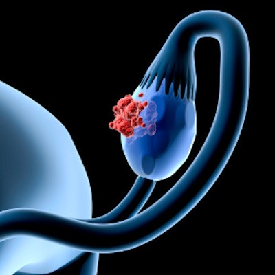
FDG-PET/CT is more accurate than contrast-enhanced CT for detecting primary ovarian cancer, but due to CT's wide availability, cost-effectiveness, and quick interpretation by junior radiologists, it is preferred on a daily basis, attendees learned at the recent World Congress on obstetrics and gynecology in London.
What the guidelines say
The European Society of Urogenital Radiology (ESUR) recommends CT for staging and in recurrent disease, and it advocates FDG-PET/CT as the "optimal imaging modality for suspected recurrences, particularly in patients with negative CT or MR findings."
On the other hand, current guidelines from the U.K. regulator, the National Institute for Health and Care Excellence (NICE), favor ultrasound and CT for diagnosing and characterizing ovarian malignancies, with there not being enough evidence for the use of FDG-PET/CT as a primary diagnostic tool.
Against this background, the researchers aimed to determine which imaging modality is most accurate in the diagnosis of primary ovarian carcinoma: FDG-PET/CT or contrast-enhanced CT. They scoured the literature for relevant articles published between January 2000 and December 2018.
Out of the 253 studies identified by the initial search, only two prospective cohort studies (Michielsen et al, European Radiology, April 2014, Vol. 24:4, pp. 889-901, 32 patients; and Dauwen H et al, Gynecologic Oncology, December 2013, Vol. 131:3, pp. 694-700, 56 patients) met the inclusion criteria, the authors told delegates at the congress, held in London from 17 to 19 June.
They analyzed a total of 88 patients, each of whom had undergone both contrast-enhanced CT and FDG-PET/CT scans or only an FDG-PET/CT scan with contrast; for the latter, each component of the scan was interpreted separately by different radiologists blinded to the other component's findings.
The key findings were as follows:
- For sensitivity, both FDG-PET/CT and contrast CT achieved a mean value of 96%, with PET/CT's sensitivity reaching 100% (Michielsen et al).
- The specificity of FDG-PET/CT ranged from 33% (Michielsen et al) to 77% (Dauwen et al), with an average value of 55%. Contrast CT's pooled specificity was significantly lower at 31% (25% Michielsen et al and 38% Dauwen et al).
- PET/CT's diagnostic accuracy was 92% (94% Michielsen et al and 90% Dauwen et al). In comparison, contrast CT's accuracy was 87%.
Overall, the Michielsen study showed that for identifying and characterizing primary ovarian tumors, FDG-PET/CT is superior to contrast CT (and similar to whole-body diffusion-weighted MRI). Meanwhile, the Dauwen study demonstrated that PET/CT can play a role as an additional technique for diagnosing ovarian cancer, the authors noted.
Due to the small number of patients in these two studies, further evidence based on a larger sample with multicenter involvement is recommended to confidently confirm these findings, they concluded.


![Examples of ultrasound findings and techniques. (A) Images in a 39-year-old male patient with a mass in the left thigh. The mass is heterogeneous on the B-mode US image (compared with the patient in D) and showed increased microvascularity (superb microvascular imaging [SMI]) and shear-wave elastography (SWE) values. Undifferentiated pleomorphic sarcoma was diagnosed at biopsy (with pleomorphic rhabdomyosarcoma in surgical specimen). (B) Images in an 18-year-old male patient with a mass in the left leg. The mass is hypoechoic on the B-mode image, with no other findings suggestive of malignancy. The lesion is in contact with the cortex of the tibia, which is slightly irregular. CT revealed a doubtful anteromedial tibial erosion. The microvascular study demonstrated high vascularization, suggestive of malignancy. Periosteal Ewing sarcoma was diagnosed with both histologic and immunohistochemical confirmation. (C) Images in a 69-year-old female patient with a lump growing on the outside of the left leg. Multiple SWE examinations were performed (please note the high values obtained in the measurements, whereas the color map highlights the stiffness relative to adjacent tissues). SMI showed areas of increased vascularization to target for sampling. Undifferentiated spindle cell sarcoma was diagnosed at biopsy, with residual leiomyosarcoma in the surgical specimen after neoadjuvant therapy. (D) Images in a 56-year-old female patient with a mass in the right thigh. The mass is heterogeneous at both B-mode ultrasound (similar to patient A) and MRI (coronal T2-weighted spectral attenuated inversion recovery [SPAIR]; T1-weighted pre-contrast and postcontrast imaging), which even shows uptake after the administration of paramagnetic contrast material, which is traditionally suggestive of malignancy. Low values at SMI and elastography are suggestive of benignity. Spindle cell lipoma was diagnosed at biopsy, with atypical spindle cell lipomatous tumor in the surgical specimen.](https://img.auntminnieeurope.com/mindful/smg/workspaces/default/uploads/2026/08/images-radiol250278fig2.APCFLSvX6p.jpg?auto=format%2Ccompress&fit=crop&h=100&q=70&w=100)


![Examples of ultrasound findings and techniques. (A) Images in a 39-year-old male patient with a mass in the left thigh. The mass is heterogeneous on the B-mode US image (compared with the patient in D) and showed increased microvascularity (superb microvascular imaging [SMI]) and shear-wave elastography (SWE) values. Undifferentiated pleomorphic sarcoma was diagnosed at biopsy (with pleomorphic rhabdomyosarcoma in surgical specimen). (B) Images in an 18-year-old male patient with a mass in the left leg. The mass is hypoechoic on the B-mode image, with no other findings suggestive of malignancy. The lesion is in contact with the cortex of the tibia, which is slightly irregular. CT revealed a doubtful anteromedial tibial erosion. The microvascular study demonstrated high vascularization, suggestive of malignancy. Periosteal Ewing sarcoma was diagnosed with both histologic and immunohistochemical confirmation. (C) Images in a 69-year-old female patient with a lump growing on the outside of the left leg. Multiple SWE examinations were performed (please note the high values obtained in the measurements, whereas the color map highlights the stiffness relative to adjacent tissues). SMI showed areas of increased vascularization to target for sampling. Undifferentiated spindle cell sarcoma was diagnosed at biopsy, with residual leiomyosarcoma in the surgical specimen after neoadjuvant therapy. (D) Images in a 56-year-old female patient with a mass in the right thigh. The mass is heterogeneous at both B-mode ultrasound (similar to patient A) and MRI (coronal T2-weighted spectral attenuated inversion recovery [SPAIR]; T1-weighted pre-contrast and postcontrast imaging), which even shows uptake after the administration of paramagnetic contrast material, which is traditionally suggestive of malignancy. Low values at SMI and elastography are suggestive of benignity. Spindle cell lipoma was diagnosed at biopsy, with atypical spindle cell lipomatous tumor in the surgical specimen.](https://img.auntminnieeurope.com/mindful/smg/workspaces/default/uploads/2026/08/images-radiol250278fig2.APCFLSvX6p.jpg?auto=format%2Ccompress&fit=crop&h=112&q=70&w=112)