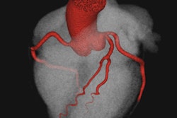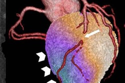
In stable patients with chest pain and suspected coronary artery disease, using coronary CT angiography (CCTA) to guide the selective use of invasive coronary angiography is safer and less expensive than sending patients directly to angiography, according to results presented on Monday at the European Society of Cardiology (ESC) congress in Rome.
The international, multicenter Coronary Computed Tomographic Angiography for Selective Cardiac Catheterization (CONSERVE) study showed that using CCTA to determine whether invasive coronary artery interventions are needed did not alter the rate of adverse cardiac events, but led to almost 90% fewer invasive procedures than direct referral to angiography.
"Our study observed lower rates of invasive procedures, which were also associated with cost savings," commented Dr. Hyuk-Jae Chang, PhD, from Yonsei University College of Medicine in Seoul, in a statement accompanying release of the results.
Nearly two-thirds of chest pain patients undergoing invasive cardiac procedures such as angiography have no actionable coronary artery disease. So whether invasive angiography will be helpful is unknown to them and their physicians.
Researchers from six countries and 18 centers examined 1,631 chest pain patients with suspected coronary artery disease. The study included patients who had undergone noninvasive stress testing, with symptoms of coronary disease, and/or preoperative evaluation for coronary artery disease. Excluded were patients with a history of coronary artery disease, interventions, or contraindications.
The primary end point of the study was major adverse cardiac events including death, nonfatal myocardial infarction, unstable angina, stroke, revascularization, or hospitalization for cerebrovascular disease. The secondary end points were economic and included within-trial costs related to coronary disease, as well as testing, hospitalization, and revascularization.
Similar patients, 2 paths
The analyzed sample included 1,503 patients randomized to either direct (n = 719) or selective (n = 794) invasive angiography for a median 12.3-month follow-up period. The two groups had no significant differences in age, gender, race, or coronary artery disease risk factors. They were also alike in angina typicality and pretest likelihood of coronary artery disease.
The results showed no statistically significant differences between the groups in rates of obstructive coronary artery disease, vessel involvement, or location of coronary disease, regardless of whether patients were sent directly to invasive coronary angiography (ICA) or sent based on CCTA results. For the primary end point of 12-month major adverse cardiac events, the rate was 5% in both groups.
For the secondary end point of mean cardiovascular cost per patient, CCTA notched substantially lower costs at $3,338 per patient, compared with sending patients directly to invasive coronary angiography, which cost $6,740 per patient. Accounting for the 4.6 million catheterizations in the U.S. each year (3.6 million in the outpatient setting), the savings come to $10 billion annually.
| Resource utilization and costs per patient | |||||
| Procedure | Direct ICA group | CCTA group | p-value | Savings with CCTA | |
| Percentage of patients receiving initial plus downstream tests | 1.02% | 0.22% | < 0.001 | 78% | |
| Percentage of patients receiving revascularization | 0.17% | 0.10% | < 0.001 | 41% | |
| Costs | $6,740 | $3,338 | < 0.001 | 60% | |
So, along with being economically meaningful, the large reduction in invasive procedures is clinically important, Chang said in the statement.
"CT-guided strategy may uncouple the diagnosis-treatment cascade of ICA which promotes excess revascularization and subsequently exposes patients to nonnegligible risk related to invasive procedure," he said. "The message from this trial is that, if we use coronary CT angiography as a gatekeeper to the catheterization lab in stable symptomatic patients with suspected coronary artery disease, we'll reduce costs with sufficient safety."


![Examples of ultrasound findings and techniques. (A) Images in a 39-year-old male patient with a mass in the left thigh. The mass is heterogeneous on the B-mode US image (compared with the patient in D) and showed increased microvascularity (superb microvascular imaging [SMI]) and shear-wave elastography (SWE) values. Undifferentiated pleomorphic sarcoma was diagnosed at biopsy (with pleomorphic rhabdomyosarcoma in surgical specimen). (B) Images in an 18-year-old male patient with a mass in the left leg. The mass is hypoechoic on the B-mode image, with no other findings suggestive of malignancy. The lesion is in contact with the cortex of the tibia, which is slightly irregular. CT revealed a doubtful anteromedial tibial erosion. The microvascular study demonstrated high vascularization, suggestive of malignancy. Periosteal Ewing sarcoma was diagnosed with both histologic and immunohistochemical confirmation. (C) Images in a 69-year-old female patient with a lump growing on the outside of the left leg. Multiple SWE examinations were performed (please note the high values obtained in the measurements, whereas the color map highlights the stiffness relative to adjacent tissues). SMI showed areas of increased vascularization to target for sampling. Undifferentiated spindle cell sarcoma was diagnosed at biopsy, with residual leiomyosarcoma in the surgical specimen after neoadjuvant therapy. (D) Images in a 56-year-old female patient with a mass in the right thigh. The mass is heterogeneous at both B-mode ultrasound (similar to patient A) and MRI (coronal T2-weighted spectral attenuated inversion recovery [SPAIR]; T1-weighted pre-contrast and postcontrast imaging), which even shows uptake after the administration of paramagnetic contrast material, which is traditionally suggestive of malignancy. Low values at SMI and elastography are suggestive of benignity. Spindle cell lipoma was diagnosed at biopsy, with atypical spindle cell lipomatous tumor in the surgical specimen.](https://img.auntminnieeurope.com/mindful/smg/workspaces/default/uploads/2026/08/images-radiol250278fig2.APCFLSvX6p.jpg?auto=format%2Ccompress&fit=crop&h=100&q=70&w=100)


![Examples of ultrasound findings and techniques. (A) Images in a 39-year-old male patient with a mass in the left thigh. The mass is heterogeneous on the B-mode US image (compared with the patient in D) and showed increased microvascularity (superb microvascular imaging [SMI]) and shear-wave elastography (SWE) values. Undifferentiated pleomorphic sarcoma was diagnosed at biopsy (with pleomorphic rhabdomyosarcoma in surgical specimen). (B) Images in an 18-year-old male patient with a mass in the left leg. The mass is hypoechoic on the B-mode image, with no other findings suggestive of malignancy. The lesion is in contact with the cortex of the tibia, which is slightly irregular. CT revealed a doubtful anteromedial tibial erosion. The microvascular study demonstrated high vascularization, suggestive of malignancy. Periosteal Ewing sarcoma was diagnosed with both histologic and immunohistochemical confirmation. (C) Images in a 69-year-old female patient with a lump growing on the outside of the left leg. Multiple SWE examinations were performed (please note the high values obtained in the measurements, whereas the color map highlights the stiffness relative to adjacent tissues). SMI showed areas of increased vascularization to target for sampling. Undifferentiated spindle cell sarcoma was diagnosed at biopsy, with residual leiomyosarcoma in the surgical specimen after neoadjuvant therapy. (D) Images in a 56-year-old female patient with a mass in the right thigh. The mass is heterogeneous at both B-mode ultrasound (similar to patient A) and MRI (coronal T2-weighted spectral attenuated inversion recovery [SPAIR]; T1-weighted pre-contrast and postcontrast imaging), which even shows uptake after the administration of paramagnetic contrast material, which is traditionally suggestive of malignancy. Low values at SMI and elastography are suggestive of benignity. Spindle cell lipoma was diagnosed at biopsy, with atypical spindle cell lipomatous tumor in the surgical specimen.](https://img.auntminnieeurope.com/mindful/smg/workspaces/default/uploads/2026/08/images-radiol250278fig2.APCFLSvX6p.jpg?auto=format%2Ccompress&fit=crop&h=112&q=70&w=112)