Dear CT Insider,
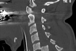
Demand is growing for emergency CT scans of the cervical spine following trauma, and because many of these examinations are performed at night or on weekends, often radiology trainees end up reporting them, according to new research from the U.K.
To speed up patient care and cut errors, the authors looked at how to improve training in this area, focusing on seven key learning points. Click here to find out more.
If an athlete at the Rio Olympics requires CT, the scan won't be conducted onsite because the organizers of the games decided against including a CT unit in the polyclinic. Presumably this decision was made due to cost and space considerations; the polyclinic in Rio occupies only 3,500 square meters, compared with 5,000 square meters at London 2012, where CT did feature. Get the full story here.
Everybody agrees CT guidance is vital for many interventional procedures, but opinion remains divided about whether interventional radiology should be a separate discipline. What's the view of Germany's new boss, Dr. Christian Stroszczynski? To learn more, click here.
European presenters excelled at the recent International Symposium on CT in San Francisco, and one of the speakers of note was Dr. Koen Nieman, PhD, from Rotterdam University Medical Center in the Netherlands. He presented cutting-edge research on the use of fractional flow reserve CT to assess the severity of coronary disease at CT angiography. Click here to read more.
Meanwhile, Willi Kalender, PhD, and his colleagues in Erlangen, Germany, have published a new study about breast CT. He's another top researcher you simply can't afford to ignore. Find out more here.
This letter outlines just a few of the many articles posted in your CT Community. For the full lineup, please check out the listing below.


![Overview of the study design. (A) The fully automated deep learning framework was developed to estimate body composition (BC) (defined as subcutaneous adipose tissue [SAT] in liters; visceral adipose tissue [VAT] in liters; skeletal muscle [SM] in liters; SM fat fraction [SMFF] as a percentage; and intramuscular adipose tissue [IMAT] in deciliters) from MRI. The fully automated framework comprised one model (model 1) to quantify different BC measures (SAT, VAT, SM, SMFF, and IMAT) as three-dimensional (3D) measures from whole-body MRI scans. The second model (model 2) was trained to identify standardized anatomic landmarks along the craniocaudal body axis (z coordinate field), which allowed for subdividing the whole-body measures into different subregions typically examined on clinical routine MRI scans (chest, abdomen, and pelvis). (B) BC was quantified from whole-body MRI in over 66,000 individuals from two large population-based cohort studies, the UK Biobank (UKB) (36,317 individuals) and the German National Cohort (NAKO) (30,291 individuals). Bar graphs show age distribution by sex and cohort. BMI = body mass index. (C) After the performance assessment of the fully automated framework, the change in BC measures, distributions, and profiles across age decades were investigated. Age-, sex-, and height-adjusted body composition reference curves were calculated and made publicly available in a web-based z-score calculator (https://circ-ml.github.io).](https://img.auntminnieeurope.com/mindful/smg/workspaces/default/uploads/2026/05/body-comp.XgAjTfPj1W.jpg?auto=format%2Ccompress&dpr=2&fit=crop&h=167&q=70&w=250)


