Dr. Maria Emília Silvestre, president of the ECR in 1987 and an important figure in Portuguese radiology, died on 18 June at the age of 84, the Portuguese Society of Radiology and Nuclear Medicine (SPRMN) has announced. She was also a prolific maker and exhibitor of patchwork quilts.
Her finest hour: Dr. Maria Emília Silvestre was president of ECR 1987. Image courtesy of SPRMN.Dr. Silvestre served the SPRMN for many years, and was president and member of the board from 1983 to 1991. In 1987, she was president of the 6th ECR, which was held in Lisbon from 31 May to 6 June 1987 and still is remembered as being a great success, according to Dr. Filipe Caseiro Alves, PhD, president of the SPRMN and professor at the University Clinic of Radiology, Coimbra University Hospitals.
"She was instrumental for Portuguese radiology, especially in the field of oncologic radiotherapy and breast cancer screening," he noted in a tribute on the SPRMN's website. "For her many roles and commitment to European radiology, she was appointed honorary member of RSNA in 1991."
Dr. Silvestre served the ECR in many functions, and was responsible for the European Diploma of Clinical Sonography from 1989 to 1992, he added.
Dr. Filipe Caseiro-Alves, PhD, from Coimbra, Portugal. Image courtesy of the ESR.
Other tributes have been paid.
"It is indeed very sad news to learn about the demise of Dr. Maria Emília Silvestre. This is a grievous loss for the Portuguese Society of Radiology and for the entire European radiological community," wrote European Society of Radiology (ESR) President Dr. Paul Parizel, PhD, in a statement posted on the SPMRN website. "On behalf of all her friends and colleagues in the ESR and ECR, I would like to express my deepest sympathies and heartfelt condolences to the bereaved family."
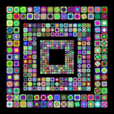
In her spare time and retirement, Dr. Silvestre was an enthusiastic creator and collector of patchwork quilts. In March-April 2014, she organized a three-week exhibition, called "Variações em Patchwork" (Variations in Patchwork).
According to the organizers of the exhibition, "The case of Maria Emília Silvestre is exemplary. After practicing medicine for 36 years, with a lot of responsibility and dedication, she turned herself in recent years to the collection, preparation, and creation of patchwork, where traditional references mix with modern decor."
Patchwork quilting remains a popular hobby in Portugal.For many years in Portugal, producing quilts and taleigos (bags) has been a popular, traditional art form. "It is that manual work, generally, that allows space for free thinking. As additional data, it is curious to note that these arts are more self-taught than taught by cultural institutions," they added.
Editor's note: A close colleague, Dr. Francisco Abecasis, has kindly agreed to write a tribute article for AuntMinnieEurope.com about Dr. Silvestre. We will publish it soon. In the meantime, if you have any personal memories of Dr. Silvestre that you would like to share, please do post them below this article.


![Examples of ultrasound findings and techniques. (A) Images in a 39-year-old male patient with a mass in the left thigh. The mass is heterogeneous on the B-mode US image (compared with the patient in D) and showed increased microvascularity (superb microvascular imaging [SMI]) and shear-wave elastography (SWE) values. Undifferentiated pleomorphic sarcoma was diagnosed at biopsy (with pleomorphic rhabdomyosarcoma in surgical specimen). (B) Images in an 18-year-old male patient with a mass in the left leg. The mass is hypoechoic on the B-mode image, with no other findings suggestive of malignancy. The lesion is in contact with the cortex of the tibia, which is slightly irregular. CT revealed a doubtful anteromedial tibial erosion. The microvascular study demonstrated high vascularization, suggestive of malignancy. Periosteal Ewing sarcoma was diagnosed with both histologic and immunohistochemical confirmation. (C) Images in a 69-year-old female patient with a lump growing on the outside of the left leg. Multiple SWE examinations were performed (please note the high values obtained in the measurements, whereas the color map highlights the stiffness relative to adjacent tissues). SMI showed areas of increased vascularization to target for sampling. Undifferentiated spindle cell sarcoma was diagnosed at biopsy, with residual leiomyosarcoma in the surgical specimen after neoadjuvant therapy. (D) Images in a 56-year-old female patient with a mass in the right thigh. The mass is heterogeneous at both B-mode ultrasound (similar to patient A) and MRI (coronal T2-weighted spectral attenuated inversion recovery [SPAIR]; T1-weighted pre-contrast and postcontrast imaging), which even shows uptake after the administration of paramagnetic contrast material, which is traditionally suggestive of malignancy. Low values at SMI and elastography are suggestive of benignity. Spindle cell lipoma was diagnosed at biopsy, with atypical spindle cell lipomatous tumor in the surgical specimen.](https://img.auntminnieeurope.com/mindful/smg/workspaces/default/uploads/2026/08/images-radiol250278fig2.APCFLSvX6p.jpg?auto=format%2Ccompress&fit=crop&h=100&q=70&w=100)


![Examples of ultrasound findings and techniques. (A) Images in a 39-year-old male patient with a mass in the left thigh. The mass is heterogeneous on the B-mode US image (compared with the patient in D) and showed increased microvascularity (superb microvascular imaging [SMI]) and shear-wave elastography (SWE) values. Undifferentiated pleomorphic sarcoma was diagnosed at biopsy (with pleomorphic rhabdomyosarcoma in surgical specimen). (B) Images in an 18-year-old male patient with a mass in the left leg. The mass is hypoechoic on the B-mode image, with no other findings suggestive of malignancy. The lesion is in contact with the cortex of the tibia, which is slightly irregular. CT revealed a doubtful anteromedial tibial erosion. The microvascular study demonstrated high vascularization, suggestive of malignancy. Periosteal Ewing sarcoma was diagnosed with both histologic and immunohistochemical confirmation. (C) Images in a 69-year-old female patient with a lump growing on the outside of the left leg. Multiple SWE examinations were performed (please note the high values obtained in the measurements, whereas the color map highlights the stiffness relative to adjacent tissues). SMI showed areas of increased vascularization to target for sampling. Undifferentiated spindle cell sarcoma was diagnosed at biopsy, with residual leiomyosarcoma in the surgical specimen after neoadjuvant therapy. (D) Images in a 56-year-old female patient with a mass in the right thigh. The mass is heterogeneous at both B-mode ultrasound (similar to patient A) and MRI (coronal T2-weighted spectral attenuated inversion recovery [SPAIR]; T1-weighted pre-contrast and postcontrast imaging), which even shows uptake after the administration of paramagnetic contrast material, which is traditionally suggestive of malignancy. Low values at SMI and elastography are suggestive of benignity. Spindle cell lipoma was diagnosed at biopsy, with atypical spindle cell lipomatous tumor in the surgical specimen.](https://img.auntminnieeurope.com/mindful/smg/workspaces/default/uploads/2026/08/images-radiol250278fig2.APCFLSvX6p.jpg?auto=format%2Ccompress&fit=crop&h=112&q=70&w=112)