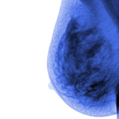
Low- and middle-income countries are seeing a rise in breast cancer incidence and death rates, despite the fact that death rates have decreased in higher-income countries, where incidence rates have increased or stabilized, according to a new study published in Cancer Epidemiology, Biomarkers and Prevention.
Although breast cancer was once considered to be primarily a disease of Western women, 52% of new cases and 62% of deaths now occur in economically developing countries, wrote the group led by Carol DeSantis of the American Cancer Society (ACS).
DeSantis and colleagues used incidence data for 39 countries from the International Agency for Research on Cancer (IARC) and mortality data for 57 countries from the World Health Organization (WHO) to assess international trends (Cancer Epidemiol Biomarkers Prev, September 10, 2015).
They found that breast cancer incidence rates are increasing in most countries, including those with historically higher rates, such as Europe, as well as in many countries in Latin America, Asia, and Africa -- regions with historically lower incidence rates. Incidence rates are stable in North America and Oceania and have declined in four European countries and Israel.
The researchers suggest that the uptick in breast cancer incidence in developing countries may be due to an increase in risk factors associated with economic development and urbanization, such as obesity, adoption of a Western-type diet, physical inactivity, delayed childbearing and having fewer children, earlier age at menarche, and shorter duration of breastfeeding.
In contrast to incidence rates, breast cancer death rates have decreased in most countries, especially high-income ones, due to improved breast cancer treatment and early detection through mammography, the group wrote. However, 10 countries are the exception to this rule: Colombia, Ecuador, Japan, Brazil, Egypt, Guatemala, Kuwait, Mauritius, Mexico, and Moldova.
"It is necessary to increase awareness about breast cancer and the benefits of early detection, most notably in economically transitioning countries, in order to successfully implement breast cancer control programs, as well as to improve access to treatment," DeSantis and colleagues concluded.


![Examples of ultrasound findings and techniques. (A) Images in a 39-year-old male patient with a mass in the left thigh. The mass is heterogeneous on the B-mode US image (compared with the patient in D) and showed increased microvascularity (superb microvascular imaging [SMI]) and shear-wave elastography (SWE) values. Undifferentiated pleomorphic sarcoma was diagnosed at biopsy (with pleomorphic rhabdomyosarcoma in surgical specimen). (B) Images in an 18-year-old male patient with a mass in the left leg. The mass is hypoechoic on the B-mode image, with no other findings suggestive of malignancy. The lesion is in contact with the cortex of the tibia, which is slightly irregular. CT revealed a doubtful anteromedial tibial erosion. The microvascular study demonstrated high vascularization, suggestive of malignancy. Periosteal Ewing sarcoma was diagnosed with both histologic and immunohistochemical confirmation. (C) Images in a 69-year-old female patient with a lump growing on the outside of the left leg. Multiple SWE examinations were performed (please note the high values obtained in the measurements, whereas the color map highlights the stiffness relative to adjacent tissues). SMI showed areas of increased vascularization to target for sampling. Undifferentiated spindle cell sarcoma was diagnosed at biopsy, with residual leiomyosarcoma in the surgical specimen after neoadjuvant therapy. (D) Images in a 56-year-old female patient with a mass in the right thigh. The mass is heterogeneous at both B-mode ultrasound (similar to patient A) and MRI (coronal T2-weighted spectral attenuated inversion recovery [SPAIR]; T1-weighted pre-contrast and postcontrast imaging), which even shows uptake after the administration of paramagnetic contrast material, which is traditionally suggestive of malignancy. Low values at SMI and elastography are suggestive of benignity. Spindle cell lipoma was diagnosed at biopsy, with atypical spindle cell lipomatous tumor in the surgical specimen.](https://img.auntminnieeurope.com/mindful/smg/workspaces/default/uploads/2026/08/images-radiol250278fig2.APCFLSvX6p.jpg?auto=format%2Ccompress&fit=crop&h=100&q=70&w=100)


![Examples of ultrasound findings and techniques. (A) Images in a 39-year-old male patient with a mass in the left thigh. The mass is heterogeneous on the B-mode US image (compared with the patient in D) and showed increased microvascularity (superb microvascular imaging [SMI]) and shear-wave elastography (SWE) values. Undifferentiated pleomorphic sarcoma was diagnosed at biopsy (with pleomorphic rhabdomyosarcoma in surgical specimen). (B) Images in an 18-year-old male patient with a mass in the left leg. The mass is hypoechoic on the B-mode image, with no other findings suggestive of malignancy. The lesion is in contact with the cortex of the tibia, which is slightly irregular. CT revealed a doubtful anteromedial tibial erosion. The microvascular study demonstrated high vascularization, suggestive of malignancy. Periosteal Ewing sarcoma was diagnosed with both histologic and immunohistochemical confirmation. (C) Images in a 69-year-old female patient with a lump growing on the outside of the left leg. Multiple SWE examinations were performed (please note the high values obtained in the measurements, whereas the color map highlights the stiffness relative to adjacent tissues). SMI showed areas of increased vascularization to target for sampling. Undifferentiated spindle cell sarcoma was diagnosed at biopsy, with residual leiomyosarcoma in the surgical specimen after neoadjuvant therapy. (D) Images in a 56-year-old female patient with a mass in the right thigh. The mass is heterogeneous at both B-mode ultrasound (similar to patient A) and MRI (coronal T2-weighted spectral attenuated inversion recovery [SPAIR]; T1-weighted pre-contrast and postcontrast imaging), which even shows uptake after the administration of paramagnetic contrast material, which is traditionally suggestive of malignancy. Low values at SMI and elastography are suggestive of benignity. Spindle cell lipoma was diagnosed at biopsy, with atypical spindle cell lipomatous tumor in the surgical specimen.](https://img.auntminnieeurope.com/mindful/smg/workspaces/default/uploads/2026/08/images-radiol250278fig2.APCFLSvX6p.jpg?auto=format%2Ccompress&fit=crop&h=112&q=70&w=112)