MRI scans have found that boys who faced challenges during the first six years of their lives had less cortical gray brain matter and altered brain structure by the time they reached their late teens or early 20s, according to a study conducted at a leading U.K. institution.
Researchers at King's College London used MRI to discover how childhood experiences affected the brain. Their work, published online on 17 August in JAMA Pediatrics, opens the possibility that intervention could help prevent children from developing internalizing symptoms and protect them against abnormal brain development.
Researchers led by Sarah K. G. Jensen began their prospective study of 494 mother-son pairs in 1991, when the mothers were expecting their newborns. The mothers reported on 37 adverse events that their families faced, such as interpersonal loss, family instability, and abuse toward the child and/or mother, when the children reached 8, 21, 33, 47, 61, and 73 months of age.
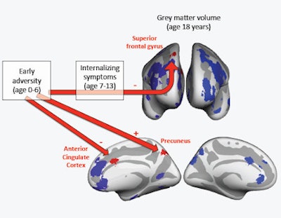
 Findings from the study by Jensen et al published in JAMA Pediatrics this week. Gray matter volumes from 494 male participants were extracted using the FreeSurfer software in 30 regions of interest, shown here on an inflated brain (top: frontal view; bottom: medial views). Regions of interest found to associate with early adversity or internalizing symptoms are shown in red, while regions of interest that did not associate with adversity or internalizing symptoms are shown in blue. "-" indicates an association with lower volume, "+" indicates an association with greater volume. Image courtesy of Sarah K. G. Jensen.
Mothers also recorded levels of internalizing symptoms, such as depression and/or anxiety, when the boys reached 7, 10, and 13 years old. At each time point, researchers created a cumulative index ranging from 0 to 37 based on the number of reported adverse events experienced by the child.
The boys were then scanned with MRI with T1-weighted imaging using a 3D fast-spoiled gradient-echo sequence acquired on a 3-tesla scanner (GE Healthcare), using an eight-channel, receiver-only head coil. Researchers measured gray-matter volume, cortical thickness, and surface area for each region of interest.
The analysis of MR images found that early adversity was directly associated with reduced gray-matter volumes in the anterior cingulate cortex (β = -0.18; p = 0.01) and higher gray-matter volume in the precuneus (β = 0.18; p = 0.009). Childhood internalizing symptoms also were associated with lower gray-matter volume in the right superior frontal gyrus (β = -0.20; p = 0.002).
(Although the study made no mention of a correlation, other previous research has linked the anterior cingulate cortex to executive functions such as memory, self-control, and awareness, and the precuneus to cognitive function.)
"The finding that childhood experiences can affect the brain highlights early childhood not only as a period of vulnerability but also as a period of opportunity," the authors wrote. "Interventions toward adversity might help to prevent children from developing internalizing symptoms and protect against abnormal brain development."
The authors noted limitations of the study, including that the cohort included only male subjects and acknowledging that other factors, other than adversity, also may affect brain development.